7SYE

Cryo-EM structure of the extracellular module of the full-length EGFR bound to EGF. "tips-separated" conformation


Experimental Data Snapshot

  • Method: ELECTRON MICROSCOPY
  • Resolution: 3.30 Å
  • Aggregation State: PARTICLE 
  • Reconstruction Method: SINGLE PARTICLE 

wwPDB Validation   3D Report Full Report


This is version 1.0 of the entry. See complete history


Literature

A molecular mechanism for the generation of ligand-dependent differential outputs by the epidermal growth factor receptor.

Huang, Y.Ognjenovic, J.Karandur, D.Miller, K.Merk, A.Subramaniam, S.Kuriyan, J.

(2021) Elife 10

  • DOI: https://doi.org/10.7554/eLife.73218
  • Primary Citation of Related Structures:  
    7SYD, 7SYE, 7SZ0, 7SZ1, 7SZ5, 7SZ7

  • PubMed Abstract: 

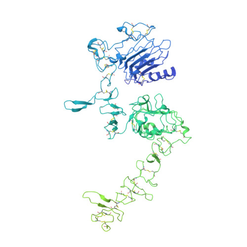
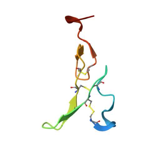
    The epidermal growth factor receptor (EGFR) is a receptor tyrosine kinase that couples the binding of extracellular ligands, such as EGF and transforming growth factor-α (TGF-α), to the initiation of intracellular signaling pathways. EGFR binds to EGF and TGF-α with similar affinity, but generates different signals from these ligands. To address the mechanistic basis of this phenomenon, we have carried out cryo-EM analyses of human EGFR bound to EGF and TGF-α. We show that the extracellular module adopts an ensemble of dimeric conformations when bound to either EGF or TGF-α. The two extreme states of this ensemble represent distinct ligand-bound quaternary structures in which the membrane-proximal tips of the extracellular module are either juxtaposed or separated. EGF and TGF-α differ in their ability to maintain the conformation with the membrane-proximal tips of the extracellular module separated, and this conformation is stabilized preferentially by an oncogenic EGFR mutation. Close proximity of the transmembrane helices at the junction with the extracellular module has been associated previously with increased EGFR activity. Our results show how EGFR can couple the binding of different ligands to differential modulation of this proximity, thereby suggesting a molecular mechanism for the generation of ligand-sensitive differential outputs in this receptor family.


  • Organizational Affiliation

    Department of Molecular and Cell Biology, University of California, Berkeley, Berkeley, United States.


Macromolecules
Find similar proteins by:  (by identity cutoff)  |  3D Structure
Entity ID: 1
MoleculeChains Sequence LengthOrganismDetailsImage
Epidermal growth factor receptor
A, B
1,210Homo sapiensMutation(s): 0 
Gene Names: EGFRERBBERBB1HER1
EC: 2.7.10.1
UniProt & NIH Common Fund Data Resources
Find proteins for P00533 (Homo sapiens)
Explore P00533 
Go to UniProtKB:  P00533
PHAROS:  P00533
GTEx:  ENSG00000146648 
Entity Groups  
Sequence Clusters30% Identity50% Identity70% Identity90% Identity95% Identity100% Identity
UniProt GroupP00533
Sequence Annotations
Expand
  • Reference Sequence
Find similar proteins by:  (by identity cutoff)  |  3D Structure
Entity ID: 2
MoleculeChains Sequence LengthOrganismDetailsImage
Epidermal growth factor
C, D
53Homo sapiensMutation(s): 0 
Gene Names: EGF
UniProt & NIH Common Fund Data Resources
Find proteins for P01133 (Homo sapiens)
Explore P01133 
Go to UniProtKB:  P01133
PHAROS:  P01133
GTEx:  ENSG00000138798 
Entity Groups  
Sequence Clusters30% Identity50% Identity70% Identity90% Identity95% Identity100% Identity
UniProt GroupP01133
Sequence Annotations
Expand
  • Reference Sequence
Experimental Data & Validation

Experimental Data

  • Method: ELECTRON MICROSCOPY
  • Resolution: 3.30 Å
  • Aggregation State: PARTICLE 
  • Reconstruction Method: SINGLE PARTICLE 
EM Software:
TaskSoftware PackageVersion
RECONSTRUCTIONcryoSPARCv2

Structure Validation

View Full Validation Report


Entry History & Funding Information

Deposition Data


Funding OrganizationLocationGrant Number
Canada Excellence Research Chair AwardUnited States--
Howard Hughes Medical Institute (HHMI)United States--

Revision History  (Full details and data files)

  • Version 1.0: 2021-12-22
    Type: Initial release