Unexpected plasticity of the quaternary structure of iron-manganese superoxide dismutases.
Mendoza Rengifo, E., Stelmastchuk Benassi Fontolan, L., Ribamar Ferreira-Junior, J., Bleicher, L., Penner-Hahn, J., Charles Garratt, R.(2022) J Struct Biol 214: 107855-107855
- PubMed: 35390463 Search on PubMed
- DOI: https://doi.org/10.1016/j.jsb.2022.107855
- Primary Citation Related Structures:
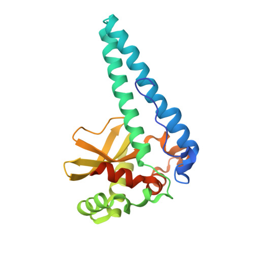
7SVS - PubMed Abstract:
Protein 3D structure can be remarkably robust to the accumulation of mutations during evolution. On the other hand, sometimes a single amino acid substitution can be sufficient to generate dramatic and completely unpredictable structural consequences. In an attempt to rationally alter the preferences for the metal ion at the active site of a member of the Iron/Manganese superoxide dismutase family, two examples of the latter phenomenon were identified. Site directed mutants of SOD from Trichoderma reesei were generated and studied crystallographically together with the wild type enzyme. Despite being chosen for their potential impact on the redox potential of the metal, two of the mutations (D150G and G73A) in fact resulted in significant alterations to the protein quaternary structure. The D150G mutant presented alternative inter-subunit contacts leading to a loss of symmetry of the wild type tetramer, whereas the G73A mutation transformed the tetramer into an octamer despite not participating directly in any of the inter-subunit interfaces. We conclude that there is considerable intrinsic plasticity in the Fe/MnSOD fold that can be unpredictably affected by single amino acid substitutions. In much the same way as phenotypic defects at the organism level can reveal much about normal function, so too can such mutations teach us much about the subtleties of protein structure.
- Laboratory of Structural Biology, Sao Carlos Institute of Physics, University of Sao Paulo, Sao Carlos, Sao Paulo, Brazil.
Organizational Affiliation: