Crystal structure of a eukaryotic phosphate transporter.
Pedersen, B.P., Kumar, H., Waight, A.B., Risenmay, A.J., Roe-Zurz, Z., Chau, B.H., Schlessinger, A., Bonomi, M., Harries, W., Sali, A., Johri, A.K., Stroud, R.M.(2013) Nature 496: 533-536
- PubMed: 23542591 Search on PubMedSearch on PubMed Central
- DOI: https://doi.org/10.1038/nature12042
- Primary Citation Related Structures:
7SP5 - PubMed Abstract:
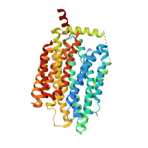
Phosphate is crucial for structural and metabolic needs, including nucleotide and lipid synthesis, signalling and chemical energy storage. Proton-coupled transporters of the major facilitator superfamily (MFS) are essential for phosphate uptake in plants and fungi, and also have a function in sensing external phosphate levels as transceptors. Here we report the 2.9 Å structure of a fungal (Piriformospora indica) high-affinity phosphate transporter, PiPT, in an inward-facing occluded state, with bound phosphate visible in the membrane-buried binding site. The structure indicates both proton and phosphate exit pathways and suggests a modified asymmetrical 'rocker-switch' mechanism of phosphate transport. PiPT is related to several human transporter families, most notably the organic cation and anion transporters of the solute carrier family (SLC22), which are implicated in cancer-drug resistance. We modelled representative cation and anion SLC22 transporters based on the PiPT structure to surmise the structural basis for substrate binding and charge selectivity in this important family. The PiPT structure demonstrates and expands on principles of substrate transport by the MFS transporters and illuminates principles of phosphate uptake in particular.
- Department of Biochemistry and Biophysics, University of California, San Francisco, California 94158, USA.
Organizational Affiliation: