7SNV

H. neapolitanus carboxysomal rubisco/CsoSCA-peptide (1-50)complex


Experimental Data Snapshot

  • Method: ELECTRON MICROSCOPY
  • Resolution: 2.07 Å
  • Aggregation State: PARTICLE 
  • Reconstruction Method: SINGLE PARTICLE 

wwPDB Validation   3D Report Full Report


This is version 1.1 of the entry. See complete history


Literature

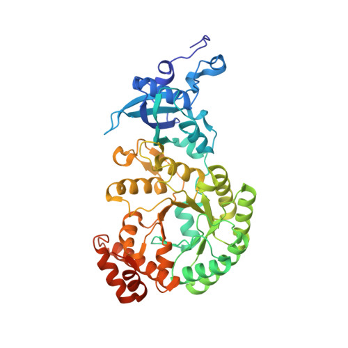
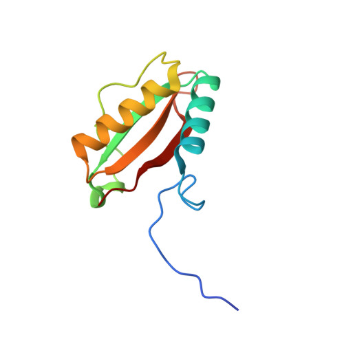
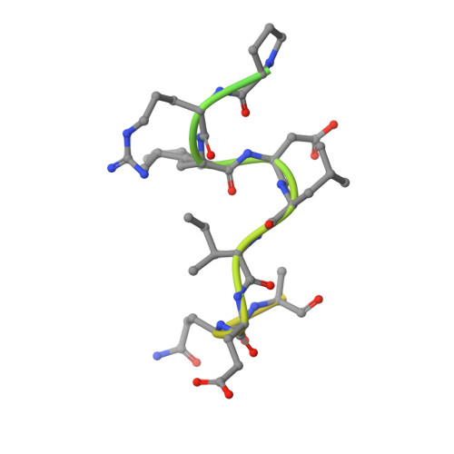
Identification of a carbonic anhydrase-Rubisco complex within the alpha-carboxysome.

Blikstad, C.Dugan, E.J.Laughlin, T.G.Turnsek, J.B.Liu, M.D.Shoemaker, S.R.Vogiatzi, N.Remis, J.P.Savage, D.F.

(2023) Proc Natl Acad Sci U S A 120: e2308600120-e2308600120

  • DOI: https://doi.org/10.1073/pnas.2308600120
  • Primary Citation of Related Structures:  
    7SMK, 7SNV

  • PubMed Abstract: 

    Carboxysomes are proteinaceous organelles that encapsulate key enzymes of CO 2 fixation-Rubisco and carbonic anhydrase-and are the centerpiece of the bacterial CO 2 concentrating mechanism (CCM). In the CCM, actively accumulated cytosolic bicarbonate diffuses into the carboxysome and is converted to CO 2 by carbonic anhydrase, producing a high CO 2 concentration near Rubisco and ensuring efficient carboxylation. Self-assembly of the α-carboxysome is orchestrated by the intrinsically disordered scaffolding protein, CsoS2, which interacts with both Rubisco and carboxysomal shell proteins, but it is unknown how the carbonic anhydrase, CsoSCA, is incorporated into the α-carboxysome. Here, we present the structural basis of carbonic anhydrase encapsulation into α-carboxysomes from Halothiobacillus neapolitanus . We find that CsoSCA interacts directly with Rubisco via an intrinsically disordered N-terminal domain. A 1.98 Å single-particle cryoelectron microscopy structure of Rubisco in complex with this peptide reveals that CsoSCA binding is predominantly mediated by a network of hydrogen bonds. CsoSCA's binding site overlaps with that of CsoS2, but the two proteins utilize substantially different motifs and modes of binding, revealing a plasticity of the Rubisco binding site. Our results advance the understanding of carboxysome biogenesis and highlight the importance of Rubisco, not only as an enzyme but also as a central hub for mediating assembly through protein interactions.


  • Organizational Affiliation

    Department of Molecular and Cell Biology, University of California, Berkeley, CA 94720.


Macromolecules
Find similar proteins by:  (by identity cutoff)  |  3D Structure
Entity ID: 1
MoleculeChains Sequence LengthOrganismDetailsImage
Ribulose bisphosphate carboxylase large chain482Halothiobacillus neapolitanus c2Mutation(s): 0 
Gene Names: cbbLHneap_0922
EC: 4.1.1.39
UniProt
Find proteins for O85040 (Halothiobacillus neapolitanus (strain ATCC 23641 / c2))
Explore O85040 
Go to UniProtKB:  O85040
Entity Groups  
Sequence Clusters30% Identity50% Identity70% Identity90% Identity95% Identity100% Identity
UniProt GroupO85040
Sequence Annotations
Expand
  • Reference Sequence
Find similar proteins by:  (by identity cutoff)  |  3D Structure
Entity ID: 2
MoleculeChains Sequence LengthOrganismDetailsImage
Ribulose bisphosphate carboxylase small chain110Halothiobacillus neapolitanus c2Mutation(s): 0 
Gene Names: cbbSrbcSHneap_0921
EC: 4.1.1.39
UniProt
Find proteins for P45686 (Halothiobacillus neapolitanus (strain ATCC 23641 / c2))
Explore P45686 
Go to UniProtKB:  P45686
Entity Groups  
Sequence Clusters30% Identity50% Identity70% Identity90% Identity95% Identity100% Identity
UniProt GroupP45686
Sequence Annotations
Expand
  • Reference Sequence
Find similar proteins by:  (by identity cutoff)  |  3D Structure
Entity ID: 3
MoleculeChains Sequence LengthOrganismDetailsImage
Carboxysome shell carbonic anhydrase50Halothiobacillus neapolitanus c2Mutation(s): 0 
EC: 4.2.1.1
UniProt
Find proteins for O85042 (Halothiobacillus neapolitanus (strain ATCC 23641 / c2))
Explore O85042 
Go to UniProtKB:  O85042
Entity Groups  
Sequence Clusters30% Identity50% Identity70% Identity90% Identity95% Identity100% Identity
UniProt GroupO85042
Sequence Annotations
Expand
  • Reference Sequence
Experimental Data & Validation

Experimental Data

  • Method: ELECTRON MICROSCOPY
  • Resolution: 2.07 Å
  • Aggregation State: PARTICLE 
  • Reconstruction Method: SINGLE PARTICLE 
EM Software:
TaskSoftware PackageVersion
MODEL REFINEMENTPHENIX1.19
RECONSTRUCTIONRELION3.1

Structure Validation

View Full Validation Report


Entry History & Funding Information

Deposition Data


Funding OrganizationLocationGrant Number
Department of Energy (DOE, United States)United StatesDE-SC00016240

Revision History  (Full details and data files)

  • Version 1.0: 2023-05-03
    Type: Initial release
  • Version 1.1: 2023-11-01
    Changes: Data collection, Database references