7SNQ | pdb_00007snq

Hexamer HIV-1 CA in complex with CPSF6 peptide and IP6 ligand


Experimental Data Snapshot

  • Method: X-RAY DIFFRACTION
  • Resolution: 2.81 Å
  • R-Value Free: 
    0.282 (Depositor), 0.276 (DCC) 
  • R-Value Work: 
    0.229 (Depositor), 0.226 (DCC) 
  • R-Value Observed: 
    0.232 (Depositor) 

Starting Model: experimental
View more details

wwPDB Validation 3D Report Full Report

Validation slider image for 7SNQ

Ligand Structure Quality Assessment 


This is version 1.3 of the entry. See complete history

Literature

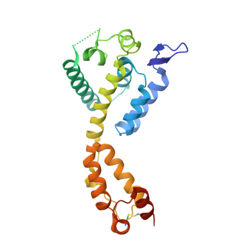
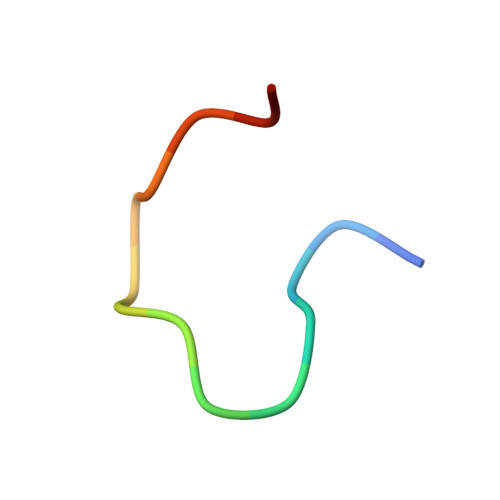
Prion-like low complexity regions enable avid virus-host interactions during HIV-1 infection.

Wei, G.Iqbal, N.Courouble, V.V.Francis, A.C.Singh, P.K.Hudait, A.Annamalai, A.S.Bester, S.Huang, S.W.Shkriabai, N.Briganti, L.Haney, R.KewalRamani, V.N.Voth, G.A.Engelman, A.N.Melikyan, G.B.Griffin, P.R.Asturias, F.Kvaratskhelia, M.

(2022) Nat Commun 13: 5879-5879

  • DOI: https://doi.org/10.1038/s41467-022-33662-6
  • Primary Citation Related Structures: 
    7SNQ

  • PubMed Abstract: 

    Cellular proteins CPSF6, NUP153 and SEC24C play crucial roles in HIV-1 infection. While weak interactions of short phenylalanine-glycine (FG) containing peptides with isolated capsid hexamers have been characterized, how these cellular factors functionally engage with biologically relevant mature HIV-1 capsid lattices is unknown. Here we show that prion-like low complexity regions (LCRs) enable avid CPSF6, NUP153 and SEC24C binding to capsid lattices. Structural studies revealed that multivalent CPSF6 assembly is mediated by LCR-LCR interactions, which are templated by binding of CPSF6 FG peptides to a subset of hydrophobic capsid pockets positioned along adjoining hexamers. In infected cells, avid CPSF6 LCR-mediated binding to HIV-1 cores is essential for functional virus-host interactions. The investigational drug lenacapavir accesses unoccupied hydrophobic pockets in the complex to potently impair HIV-1 inside the nucleus without displacing the tightly bound cellular cofactor from virus cores. These results establish previously undescribed mechanisms of virus-host interactions and antiviral action.


  • Organizational Affiliation
    • Division of Infectious Diseases, Anschutz Medical Campus, University of Colorado School of Medicine, Aurora, CO, 80045, USA.

Macromolecule Content 

  • Total Structure Weight: 326.86 kDa 
  • Atom Count: 20,569 
  • Modeled Residue Count: 2,653 
  • Deposited Residue Count: 2,952 
  • Unique protein chains: 2

Macromolecules

Find similar proteins by:|  3D Structure
Entity ID: 1
MoleculeChains  Sequence LengthOrganismDetailsImage
Capsid protein p24
A, B, C, D, E
A, B, C, D, E, F, G, H, I, J, K, L
231Human immunodeficiency virus type 1 BH10Mutation(s): 4 
Gene Names: gag-pol
UniProt
Find proteins for P03366 (Human immunodeficiency virus type 1 group M subtype B (isolate BH10))
Explore P03366 
Go to UniProtKB:  P03366
Entity Groups
Sequence Clusters30% Identity50% Identity70% Identity90% Identity95% Identity100% Identity
UniProt GroupP03366
Sequence Annotations
Expand
Reference Sequence
Find similar proteins by:  Sequence   |   3D Structure  
Entity ID: 2
MoleculeChains  Sequence LengthOrganismDetailsImage
Cleavage and polyadenylation specificity factor subunit 6
M, N, O, P, Q
M, N, O, P, Q, R, S, T, U, V, W, X
15Human immunodeficiency virus 1Mutation(s): 0 
UniProt & NIH Common Fund Data Resources
Find proteins for Q16630 (Homo sapiens)
Explore Q16630 
Go to UniProtKB:  Q16630
PHAROS:  Q16630
GTEx:  ENSG00000111605 
Entity Groups
Sequence Clusters30% Identity50% Identity70% Identity90% Identity95% Identity100% Identity
UniProt GroupQ16630
Sequence Annotations
Expand
Reference Sequence

Experimental Data & Validation

Experimental Data

  • Method: X-RAY DIFFRACTION
  • Resolution: 2.81 Å
  • R-Value Free:  0.282 (Depositor), 0.276 (DCC) 
  • R-Value Work:  0.229 (Depositor), 0.226 (DCC) 
  • R-Value Observed: 0.232 (Depositor) 
Space Group: P 21 21 21
Unit Cell:
Length ( Å )Angle ( ˚ )
a = 133.918α = 90
b = 135.673β = 90
c = 212.88γ = 90
Software Package:
Software NamePurpose
PHENIXrefinement
XDSdata reduction
XDSdata scaling
PHASERphasing

Structure Validation

View Full Validation Report


Ligand Structure Quality Assessment 


Entry History 

& Funding Information

Deposition Data


Funding OrganizationLocationGrant Number
National Institutes of Health/National Institute of General Medical Sciences (NIH/NIGMS)United StatesU54 GM103368

Revision History  (Full details and data files)

  • Version 1.0: 2022-09-07
    Type: Initial release
  • Version 1.1: 2022-10-19
    Changes: Database references
  • Version 1.2: 2023-10-18
    Changes: Data collection, Refinement description
  • Version 1.3: 2024-10-16
    Changes: Structure summary