7SH4 | pdb_00007sh4

CD1a-phosphatidylglycerol binary structure


Experimental Data Snapshot

  • Method: X-RAY DIFFRACTION
  • Resolution: 2.00 Å
  • R-Value Free: 
    0.230 (Depositor), 0.211 (DCC) 
  • R-Value Work: 
    0.193 (Depositor) 
  • R-Value Observed: 
    0.194 (Depositor) 

Starting Model: experimental
View more details

wwPDB Validation 3D Report Full Report

Validation slider image for 7SH4

Ligand Structure Quality Assessment 


This is version 1.3 of the entry. See complete history

Literature

Staphylococcal phosphatidylglycerol antigens activate human T cells via CD1a.

Monnot, G.C.Wegrecki, M.Cheng, T.Y.Chen, Y.L.Sallee, B.N.Chakravarthy, R.Karantza, I.M.Tin, S.Y.Khaleel, A.E.Monga, I.Uwakwe, L.N.Tillman, A.Cheng, B.Youssef, S.Ng, S.W.Shahine, A.Garcia-Vilas, J.A.Uhlemann, A.C.Bordone, L.A.Han, A.Rohde, C.H.Ogg, G.Moody, D.B.Rossjohn, J.de Jong, A.

(2023) Nat Immunol 24: 110-122

  • DOI: https://doi.org/10.1038/s41590-022-01375-z
  • Primary Citation Related Structures: 
    7SH4

  • PubMed Abstract: 

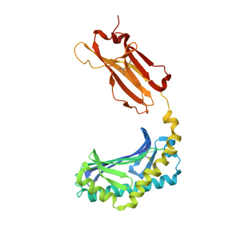
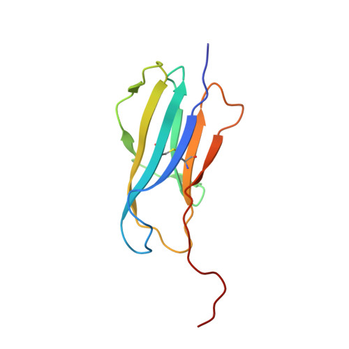
    Expressed on epidermal Langerhans cells, CD1a presents a range of self-lipid antigens found within the skin; however, the extent to which CD1a presents microbial ligands from bacteria colonizing the skin is unclear. Here we identified CD1a-dependent T cell responses to phosphatidylglycerol (PG), a ubiquitous bacterial membrane phospholipid, as well as to lysylPG, a modified PG, present in several Gram-positive bacteria and highly abundant in Staphylococcus aureus. The crystal structure of the CD1a-PG complex showed that the acyl chains were buried within the A'- and F'-pockets of CD1a, while the phosphoglycerol headgroup remained solvent exposed in the F'-portal and was available for T cell receptor contact. Using lysylPG and PG-loaded CD1a tetramers, we identified T cells in peripheral blood and in skin that respond to these lipids in a dose-dependent manner. Tetramer + CD4 +  T cell lines secreted type 2 helper T cell cytokines in response to phosphatidylglycerols as well as to co-cultures of CD1a +  dendritic cells and Staphylococcus bacteria. The expansion in patients with atopic dermatitis of CD4 + CD1a-(lysyl)PG tetramer + T cells suggests a response to lipids made by bacteria associated with atopic dermatitis and provides a link supporting involvement of PG-based lipid-activated T cells in atopic dermatitis pathogenesis.


  • Organizational Affiliation
    • Department of Dermatology, Columbia University Irving Medical Center, New York, NY, USA.

Macromolecule Content 

  • Total Structure Weight: 46.55 kDa 
  • Atom Count: 3,469 
  • Modeled Residue Count: 378 
  • Deposited Residue Count: 393 
  • Unique protein chains: 2

Macromolecules

Find similar proteins by:|  3D Structure
Entity ID: 1
MoleculeChains  Sequence LengthOrganismDetailsImage
T-cell surface glycoprotein CD1a285Homo sapiensMutation(s): 0 
Gene Names: CD1A
UniProt & NIH Common Fund Data Resources
Find proteins for P06126 (Homo sapiens)
Explore P06126 
Go to UniProtKB:  P06126
PHAROS:  P06126
GTEx:  ENSG00000158477 
Entity Groups
Sequence Clusters30% Identity50% Identity70% Identity90% Identity95% Identity100% Identity
UniProt GroupP06126
Glycosylation
Glycosylation Sites: 1Go to GlyGen: P06126-1
Sequence Annotations
Expand
Reference Sequence
Find similar proteins by:|  3D Structure
Entity ID: 2
MoleculeChains  Sequence LengthOrganismDetailsImage
Beta-2-microglobulin108Homo sapiensMutation(s): 0 
Gene Names: B2MCDABP0092HDCMA22P
UniProt & NIH Common Fund Data Resources
Find proteins for P61769 (Homo sapiens)
Explore P61769 
Go to UniProtKB:  P61769
PHAROS:  P61769
GTEx:  ENSG00000166710 
Entity Groups
Sequence Clusters30% Identity50% Identity70% Identity90% Identity95% Identity100% Identity
UniProt GroupP61769
Sequence Annotations
Expand
Reference Sequence

Small Molecules

Ligands 5 Unique
IDChains Name / Formula / InChI Key2D Diagram3D Interactions
MW9
(Subject of Investigation/LOI)

Query on MW9


Download:Ideal Coordinates CCD File
D [auth A](21R,24R,27S)-24,27,28-trihydroxy-18,24-dioxo-19,23,25-trioxa-24lambda~5~-phosphaoctacosan-21-yl (9Z)-octadec-9-enoate
C42 H81 O10 P
ZEFGRNLJASLRBZ-QIJYXWHJSA-N
NAG

Query on NAG


Download:Ideal Coordinates CCD File
C [auth A]2-acetamido-2-deoxy-beta-D-glucopyranose
C8 H15 N O6
OVRNDRQMDRJTHS-FMDGEEDCSA-N
MES

Query on MES


Download:Ideal Coordinates CCD File
F [auth A]2-(N-MORPHOLINO)-ETHANESULFONIC ACID
C6 H13 N O4 S
SXGZJKUKBWWHRA-UHFFFAOYSA-N
PEG

Query on PEG


Download:Ideal Coordinates CCD File
H [auth B],
I [auth B]
DI(HYDROXYETHYL)ETHER
C4 H10 O3
MTHSVFCYNBDYFN-UHFFFAOYSA-N
EDO

Query on EDO


Download:Ideal Coordinates CCD File
E [auth A],
G [auth A]
1,2-ETHANEDIOL
C2 H6 O2
LYCAIKOWRPUZTN-UHFFFAOYSA-N

Experimental Data & Validation

Experimental Data

  • Method: X-RAY DIFFRACTION
  • Resolution: 2.00 Å
  • R-Value Free:  0.230 (Depositor), 0.211 (DCC) 
  • R-Value Work:  0.193 (Depositor) 
  • R-Value Observed: 0.194 (Depositor) 
Space Group: P 21 21 21
Unit Cell:
Length ( Å )Angle ( ˚ )
a = 42.225α = 90
b = 89.79β = 90
c = 105.16γ = 90
Software Package:
Software NamePurpose
PHENIXrefinement
XDSdata reduction
Aimlessdata scaling
PHASERphasing

Structure Validation

View Full Validation Report


Ligand Structure Quality Assessment 


Entry History 

& Funding Information

Deposition Data


Funding OrganizationLocationGrant Number
National Health and Medical Research Council (NHMRC, Australia)Australia--

Revision History  (Full details and data files)

  • Version 1.0: 2022-10-05
    Type: Initial release
  • Version 1.1: 2023-01-18
    Changes: Database references
  • Version 1.2: 2023-10-25
    Changes: Data collection, Refinement description
  • Version 1.3: 2024-10-16
    Changes: Structure summary