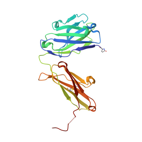
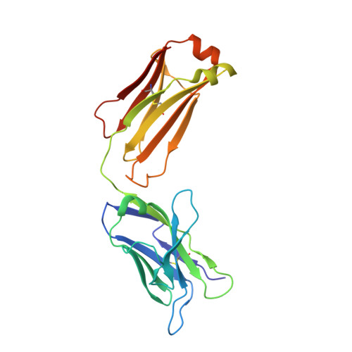
7SEN

Crystal structure of Fab containing a fluorescent noncanonical amino acid with blocked excited state proton transfer


Experimental Data Snapshot

  • Method: X-RAY DIFFRACTION
  • Resolution: 2.09 Å
  • R-Value Free: 0.253 
  • R-Value Work: 0.185 
  • R-Value Observed: 0.189 

wwPDB Validation   3D Report Full Report


This is version 1.3 of the entry. See complete history


Literature

Structural Basis for Blocked Excited State Proton Transfer in a Fluorescent, Photoacidic Non-Canonical Amino Acid-Containing Antibody Fragment.

Henderson, J.N.Simmons, C.R.Mills, J.H.

(2022) J Mol Biol 434: 167455-167455

  • DOI: https://doi.org/10.1016/j.jmb.2022.167455
  • Primary Citation of Related Structures:  
    7SEN, 7SGM

  • PubMed Abstract: 

    The fluorescent non-canonical amino acid (fNCAA) L-(7-hydroxycoumarin-4-yl)ethylglycine (7-HCAA) contains a photoacidic 7-hydroxycoumarin (7-HC) side chain whose fluorescence properties can be tuned by its environment. In proteins, many alterations to 7-HCAA's fluorescence spectra have been reported including increases and decreases in intensity and red- and blue-shifted emission maxima. The ability to rationally design protein environments that alter 7-HCAA's fluorescence properties in predictable ways could lead to novel protein-based sensors of biological function. However, these efforts are likely limited by a lack of structural characterization of 7-HCAA-containing proteins. Here, we report the steady-state spectroscopic and x-ray crystallographic characterization of a 7-HCAA-containing antibody fragment (in the apo and antigen-bound forms) in which a substantially blue-shifted 7-HCAA emission maximum (∼70 nm) is observed relative to the free amino acid. Our structural characterization of these proteins provides evidence that the blue shift is a consequence of the fact that excited state proton transfer (ESPT) from the 7-HC phenol has been almost completely blocked by interactions with the protein backbone. Furthermore, a direct interaction between a residue in the antigen and the fluorophore served to further block proton transfer relative to the apoprotein. The structural basis of the unprecedented blue shift in 7-HCAA emission reported here provides a framework for the development of new fluorescent protein-based sensors.


  • Organizational Affiliation

    The Biodesign Center for Molecular Design and Biomimetics, Arizona State University, Tempe, AZ, USA.


Macromolecules
Find similar proteins by:  (by identity cutoff)  |  3D Structure
Entity ID: 1
MoleculeChains Sequence LengthOrganismDetailsImage
5c8* Fab heavy chainA [auth H]226Homo sapiensMutation(s): 0 
Entity Groups  
Sequence Clusters30% Identity50% Identity70% Identity90% Identity95% Identity100% Identity
Sequence Annotations
Expand
  • Reference Sequence
Find similar proteins by:  (by identity cutoff)  |  3D Structure
Entity ID: 2
MoleculeChains Sequence LengthOrganismDetailsImage
5c8* Fab light chainB [auth L]218Homo sapiensMutation(s): 0 
Entity Groups  
Sequence Clusters30% Identity50% Identity70% Identity90% Identity95% Identity100% Identity
Sequence Annotations
Expand
  • Reference Sequence
Small Molecules
Modified Residues  1 Unique
IDChains TypeFormula2D DiagramParent
PCA
Query on PCA
A [auth H]L-PEPTIDE LINKINGC5 H7 N O3GLN
Experimental Data & Validation

Experimental Data

  • Method: X-RAY DIFFRACTION
  • Resolution: 2.09 Å
  • R-Value Free: 0.253 
  • R-Value Work: 0.185 
  • R-Value Observed: 0.189 
  • Space Group: C 1 2 1
Unit Cell:
Length ( Å )Angle ( ˚ )
a = 90.174α = 90
b = 59.811β = 96.09
c = 73.043γ = 90
Software Package:
Software NamePurpose
HKL-2000data scaling
PHASERphasing
REFMACrefinement
PDB_EXTRACTdata extraction
HKL-2000data reduction

Structure Validation

View Full Validation Report


Entry History & Funding Information

Deposition Data


Funding OrganizationLocationGrant Number
National Institutes of Health/National Institute of General Medical Sciences (NIH/NIGMS)United StatesR01 GM136996

Revision History  (Full details and data files)

  • Version 1.0: 2022-02-02
    Type: Initial release
  • Version 1.1: 2022-04-27
    Changes: Database references
  • Version 1.2: 2023-10-18
    Changes: Data collection, Refinement description
  • Version 1.3: 2023-11-15
    Changes: Data collection, Derived calculations