Differences in the dynamics of the tandem-SH2 modules of the Syk and ZAP-70 tyrosine kinases.
Hobbs, H.T., Shah, N.H., Badroos, J.M., Gee, C.L., Marqusee, S., Kuriyan, J.(2021) Protein Sci 30: 2373-2384
- PubMed: 34601763 Search on PubMedSearch on PubMed Central
- DOI: https://doi.org/10.1002/pro.4199
- Primary Citation Related Structures:
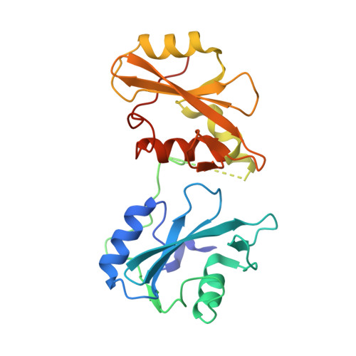
7SA7 - PubMed Abstract:
The catalytic activity of Syk-family tyrosine kinases is regulated by a tandem Src homology 2 module (tSH2 module). In the autoinhibited state, this module adopts a conformation that stabilizes an inactive conformation of the kinase domain. The binding of the tSH2 module to phosphorylated immunoreceptor tyrosine-based activation motifs necessitates a conformational change, thereby relieving kinase inhibition and promoting activation. We determined the crystal structure of the isolated tSH2 module of Syk and find, in contrast to ZAP-70, that its conformation more closely resembles that of the peptide-bound state, rather than the autoinhibited state. Hydrogen-deuterium exchange by mass spectrometry, as well as molecular dynamics simulations, reveal that the dynamics of the tSH2 modules of Syk and ZAP-70 differ, with most of these differences occurring in the C-terminal SH2 domain. Our data suggest that the conformational landscapes of the tSH2 modules in Syk and ZAP-70 have been tuned differently, such that the autoinhibited conformation of the Syk tSH2 module is less stable. This feature of Syk likely contributes to its ability to more readily escape autoinhibition when compared to ZAP-70, consistent with tighter control of downstream signaling pathways in T cells.
- Department of Chemistry, University of California, Berkeley, California, USA.
Organizational Affiliation: