The Ephb2 Receptor Uses Homotypic, Head-to-Tail Interactions within Its Ectodomain as an Autoinhibitory Control Mechanism.
Xu, Y., Robev, D., Saha, N., Wang, B., Dalva, M.B., Xu, K., Himanen, J.P., Nikolov, D.B.(2021) Int J Mol Sci 22
- PubMed: 34638814 Search on PubMedSearch on PubMed Central
- DOI: https://doi.org/10.3390/ijms221910473
- Primary Citation Related Structures:
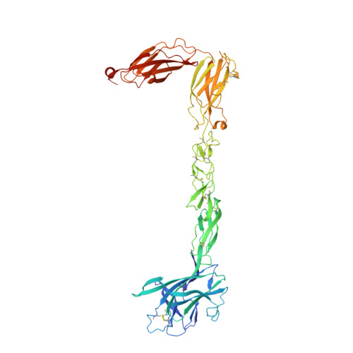
7S7K - PubMed Abstract:
The Eph receptor tyrosine kinases and their ephrin ligands direct axon pathfinding and neuronal cell migration, as well as mediate many other cell-cell communication events. Their dysfunctional signaling has been shown to lead to various diseases, including cancer. The Ephs and ephrins both localize to the plasma membrane and, upon cell-cell contact, form extensive signaling assemblies at the contact sites. The Ephs and the ephrins are divided into A and B subclasses based on their sequence conservation and affinities for each other. The molecular details of Eph-ephrin recognition have been previously revealed and it has been documented that ephrin binding induces higher-order Eph assemblies, which are essential for full biological activity, via multiple, distinct Eph-Eph interfaces. One Eph-Eph interface type is characterized by a homotypic, head-to-tail interaction between the ligand-binding and the fibronectin domains of two adjacent Eph molecules. While the previous Eph ectodomain structural studies were focused on A class receptors, we now report the crystal structure of the full ectodomain of EphB2, revealing distinct and unique head-to-tail receptor-receptor interactions. The EphB2 structure and structure-based mutagenesis document that EphB2 uses the head-to-tail interactions as a novel autoinhibitory control mechanism for regulating downstream signaling and that these interactions can be modulated by posttranslational modifications.
- Department of Veterinary Biosciences, College of Veterinary Medicine, The Ohio State University, Columbus, OH 43210, USA.
Organizational Affiliation: