Stable binding to phosphatidylserine-containing membranes requires conserved arginine residues in tandem C domains of blood coagulation factor VIII.
Peters, S.C., Childers, K.C., Mitchell, C.E., Avery, N.G., Reese Jr., S.S., Mitchell, C., Wo, S.W., Swanson, C.D., Brison, C.M., Spiegel Jr., P.C.(2022) Front Mol Biosci 9: 1040106-1040106
- PubMed: 36387287 Search on PubMedSearch on PubMed Central
- DOI: https://doi.org/10.3389/fmolb.2022.1040106
- Primary Citation Related Structures:
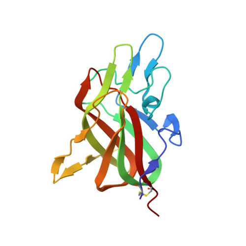
7S0P - PubMed Abstract:
At sites of vascular damage, factor VIII (fVIII) is proteolytically activated by thrombin and binds to activated platelet surfaces with activated factor IX (fIXa) to form the intrinsic "tenase" complex. Previous structural and mutational studies of fVIII have identified the C1 and C2 domains in binding to negatively charged membrane surfaces through β-hairpin loops with solvent-exposed hydrophobic residues and a ring of positively charged basic residues. Several hemophilia A-associated mutations within the C domains are suggested to disrupt lipid binding, preventing formation of the intrinsic tenase complex. In this study, we devised a novel platform for generating recombinant C1, C2, and C1C2 domain constructs and performed mutagenesis of several charged residues proximal to the putative membrane binding region of each C domain. Binding measurements between phosphatidylserine (PS)-containing lipid membrane surfaces and fVIII C domains demonstrated an ionic strength dependence on membrane binding affinity. Mutations to basic residues adjacent to the surface-exposed hydrophobic regions of C1 and C2 differentially disrupted membrane binding, with abrogation of binding occurring for mutations to conserved arginine residues in the C1 (R2163) and C2 (R2320) domains. Lastly, we determined the X-ray crystal structure of the porcine fVIII C2 domain bound to o -phospho-L-serine, the polar headgroup of PS, which binds to a basic cleft and makes charge-charge contact with R2320. We conclude that basic clefts in the fVIII C domains bind to PS-containing membranes through conserved arginine residues via a C domain modularity, where each C domain possesses modest electrostatic-dependent affinity and tandem C domains are required for high affinity binding.
- Department of Chemistry, Western Washington University, Bellingham, WA, United States.
Organizational Affiliation: