7RX9

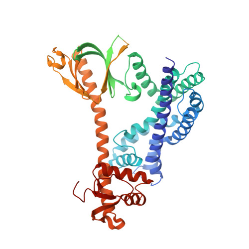
Structure of autoinhibited P-Rex1


Experimental Data Snapshot

  • Method: X-RAY DIFFRACTION
  • Resolution: 3.22 Å
  • R-Value Free: 0.262 
  • R-Value Work: 0.236 
  • R-Value Observed: 0.237 

wwPDB Validation   3D Report Full Report


This is version 1.2 of the entry. See complete history


Literature

Structure of the metastatic factor P-Rex1 reveals a two-layered autoinhibitory mechanism.

Chang, Y.G.Lupton, C.J.Bayly-Jones, C.Keen, A.C.D'Andrea, L.Lucato, C.M.Steele, J.R.Venugopal, H.Schittenhelm, R.B.Whisstock, J.C.Halls, M.L.Ellisdon, A.M.

(2022) Nat Struct Mol Biol 29: 767-773

  • DOI: https://doi.org/10.1038/s41594-022-00804-9
  • Primary Citation of Related Structures:  
    7RX9, 7SYF

  • PubMed Abstract: 

    P-Rex (PI(3,4,5)P 3 -dependent Rac exchanger) guanine nucleotide exchange factors potently activate Rho GTPases. P-Rex guanine nucleotide exchange factors are autoinhibited, synergistically activated by Gβγ and PI(3,4,5)P 3 binding and dysregulated in cancer. Here, we use X-ray crystallography, cryogenic electron microscopy and crosslinking mass spectrometry to determine the structural basis of human P-Rex1 autoinhibition. P-Rex1 has a bipartite structure of N- and C-terminal modules connected by a C-terminal four-helix bundle that binds the N-terminal Pleckstrin homology (PH) domain. In the N-terminal module, the Dbl homology (DH) domain catalytic surface is occluded by the compact arrangement of the DH-PH-DEP1 domains. Structural analysis reveals a remarkable conformational transition to release autoinhibition, requiring a 126° opening of the DH domain hinge helix. The off-axis position of Gβγ and PI(3,4,5)P 3 binding sites further suggests a counter-rotation of the P-Rex1 halves by 90° facilitates PH domain uncoupling from the four-helix bundle, releasing the autoinhibited DH domain to drive Rho GTPase signaling.


  • Organizational Affiliation

    Biomedicine Discovery Institute, Monash University, Clayton, Victoria, Australia. tyler.chang@monash.edu.


Macromolecules
Find similar proteins by:  (by identity cutoff)  |  3D Structure
Entity ID: 1
MoleculeChains Sequence LengthOrganismDetailsImage
Phosphatidylinositol 3,4,5-trisphosphate-dependent Rac exchanger 1 protein, Endolysin chimera606Homo sapiensTequatrovirus T4Mutation(s): 0 
Gene Names: PREX1KIAA1415
EC: 3.2.1.17
UniProt & NIH Common Fund Data Resources
Find proteins for Q8TCU6 (Homo sapiens)
Explore Q8TCU6 
Go to UniProtKB:  Q8TCU6
PHAROS:  Q8TCU6
GTEx:  ENSG00000124126 
Find proteins for P00720 (Enterobacteria phage T4)
Explore P00720 
Go to UniProtKB:  P00720
Entity Groups  
Sequence Clusters30% Identity50% Identity70% Identity90% Identity95% Identity100% Identity
UniProt GroupsQ8TCU6P00720
Sequence Annotations
Expand
  • Reference Sequence
Experimental Data & Validation

Experimental Data

  • Method: X-RAY DIFFRACTION
  • Resolution: 3.22 Å
  • R-Value Free: 0.262 
  • R-Value Work: 0.236 
  • R-Value Observed: 0.237 
  • Space Group: P 41 2 2
Unit Cell:
Length ( Å )Angle ( ˚ )
a = 151.256α = 90
b = 151.256β = 90
c = 94.287γ = 90
Software Package:
Software NamePurpose
PHENIXrefinement
XDSdata reduction
Aimlessdata scaling
PHASERphasing

Structure Validation

View Full Validation Report


Entry History & Funding Information

Deposition Data


Funding OrganizationLocationGrant Number
National Health and Medical Research Council (NHMRC, Australia)AustraliaAPP1128120
National Health and Medical Research Council (NHMRC, Australia)AustraliaAPP1146578

Revision History  (Full details and data files)

  • Version 1.0: 2022-08-10
    Type: Initial release
  • Version 1.1: 2022-08-24
    Changes: Database references
  • Version 1.2: 2023-10-18
    Changes: Data collection, Refinement description