Structures of the junctophilin/voltage-gated calcium channel interface reveal hot spot for cardiomyopathy mutations.
Yang, Z.F., Panwar, P., McFarlane, C.R., Tuinte, W.E., Campiglio, M., Van Petegem, F.(2022) Proc Natl Acad Sci U S A 119: e2120416119-e2120416119
- PubMed: 35238659 Search on PubMedSearch on PubMed Central
- DOI: https://doi.org/10.1073/pnas.2120416119
- Primary Citation Related Structures:
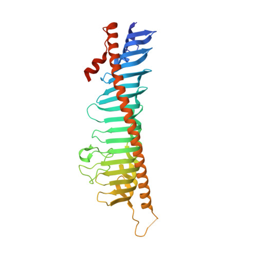
7RW4, 7RXE, 7RXQ - PubMed Abstract:
SignificanceIon channels have evolved the ability to communicate with one another, either through protein-protein interactions, or indirectly via intermediate diffusible messenger molecules. In special cases, the channels are part of different membranes. In muscle tissue, the T-tubule membrane is in proximity to the sarcoplasmic reticulum, allowing communication between L-type calcium channels and ryanodine receptors. This process is critical for excitation-contraction coupling and requires auxiliary proteins like junctophilin (JPH). JPHs are targets for disease-associated mutations, most notably hypertrophic cardiomyopathy mutations in the JPH2 isoform. Here we provide high-resolution snapshots of JPH, both alone and in complex with a calcium channel peptide, and show how this interaction is targeted by cardiomyopathy mutations.
- Department of Biochemistry and Molecular Biology, The Life Sciences Institute, University of British Columbia, Vancouver, BC V6T 1Z3, Canada.
Organizational Affiliation: