Antitarget Selectivity and Tolerability of Novel Pyrrolo[2,3- d ]pyrimidine RET Inhibitors.
Mathison, C.J.N., Yang, Y., Nelson, J., Huang, Z., Jiang, J., Chianelli, D., Rucker, P.V., Roland, J., Xie, Y.F., Epple, R., Bursulaya, B., Lee, C., Gao, M.Y., Shaffer, J., Briones, S., Sarkisova, Y., Galkin, A., Li, L., Li, N., Li, C., Hua, S., Kasibhatla, S., Kinyamu-Akunda, J., Kikkawa, R., Molteni, V., Tellew, J.E.(2021) ACS Med Chem Lett 12: 1912-1919
- PubMed: 34917254 Search on PubMedSearch on PubMed Central
- DOI: https://doi.org/10.1021/acsmedchemlett.1c00450
- Primary Citation Related Structures:
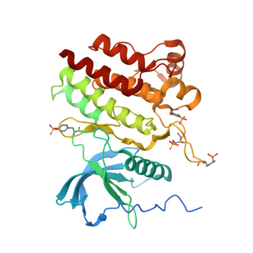
7RUN - PubMed Abstract:
The selective inhibition of RET kinase as a treatment for relevant cancer types including lung adenocarcinoma has garnered considerable interest in recent years and prompted a variety of efforts toward the discovery of small-molecule therapeutics. Hits uncovered via the analysis of archival kinase data ultimately led to the identification of a promising pyrrolo[2,3- d ]pyrimidine scaffold. The optimization of this pyrrolo[2,3- d ]pyrimidine core resulted in compound 1 , which demonstrated potent in vitro RET kinase inhibition and robust in vivo efficacy in RET-driven tumor xenografts upon multiday dosing in mice. The administration of 1 was well-tolerated at established efficacious doses (10 and 30 mg/kg, po, qd), and plasma exposure levels indicated a minimal risk of KDR or hERG inhibition in vivo , as evaluated by Miles assay and free plasma concentrations, respectively.
- The Genomics Institute of the Novartis Research Foundation, 10675 John Jay Hopkins Drive, San Diego, California 92121, United States.
Organizational Affiliation: