7RUE

DAHP synthase complexed with trifluoropyruvate semicarbazone


Experimental Data Snapshot

  • Method: X-RAY DIFFRACTION
  • Resolution: 2.50 Å
  • R-Value Free: 0.263 
  • R-Value Work: 0.201 
  • R-Value Observed: 0.204 

wwPDB Validation   3D Report Full Report


Ligand Structure Quality Assessment 


This is version 1.3 of the entry. See complete history


Literature

An Inhibitor-in-Pieces Approach to DAHP Synthase Inhibition: Potent Enzyme and Bacterial Growth Inhibition.

Heimhalt, M.Mukherjee, P.Grainger, R.A.Szabla, R.Brown, C.Turner, R.Junop, M.S.Berti, P.J.

(2021) ACS Infect Dis 7: 3292-3302

  • DOI: https://doi.org/10.1021/acsinfecdis.1c00462
  • Primary Citation of Related Structures:  
    7RUD, 7RUE

  • PubMed Abstract: 

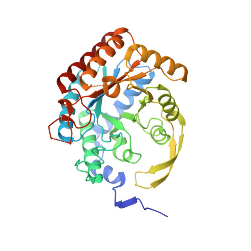
    3-Deoxy-d- arabino heptulosonate-7-phosphate (DAHP) synthase catalyzes the first step in the shikimate biosynthetic pathway and is an antimicrobial target. We used an inhibitor-in-pieces approach, based on the previously reported inhibitor DAHP oxime, to screen inhibitor fragments in the presence and absence of glycerol 3-phosphate to occupy the distal end of the active site. This led to DAHP hydrazone, the most potent inhibitor to date, K i = 10 ± 1 nM. Three trifluoropyruvate (TFP)-based inhibitor fragments were efficient inhibitors with ligand efficiencies of up to 0.7 kcal mol -1 /atom compared with 0.2 kcal mol -1 /atom for a typical good inhibitor. The crystal structures showed the TFP-based inhibitors binding upside down in the active site relative to DAHP oxime, providing new avenues for inhibitor development. The ethyl esters of TFP oxime and TFP semicarbazone prevented E. coli growth in culture with IC 50 = 0.21 ± 0.01 and 0.77 ± 0.08 mg mL -1 , respectively. Overexpressing DAHP synthase relieved growth inhibition, demonstrating that DAHP synthase was the target. Growth inhibition occurred in media containing aromatic amino acids, suggesting that growth inhibition was due to depletion of some other product(s) of the shikimate pathway, possibly folate.


  • Organizational Affiliation

    Department of Biochemistry, Molecular Biology Lab, Western University, London, Ontario N6A 5C1, Canada.


Macromolecules
Find similar proteins by:  (by identity cutoff)  |  3D Structure
Entity ID: 1
MoleculeChains Sequence LengthOrganismDetailsImage
Phospho-2-dehydro-3-deoxyheptonate aldolase, Phe-sensitive
A, B, C, D
351Escherichia coli K-12Mutation(s): 0 
Gene Names: aroGb0754JW0737
EC: 2.5.1.54
UniProt
Find proteins for P0AB91 (Escherichia coli (strain K12))
Explore P0AB91 
Go to UniProtKB:  P0AB91
Entity Groups  
Sequence Clusters30% Identity50% Identity70% Identity90% Identity95% Identity100% Identity
UniProt GroupP0AB91
Sequence Annotations
Expand
  • Reference Sequence
Experimental Data & Validation

Experimental Data

  • Method: X-RAY DIFFRACTION
  • Resolution: 2.50 Å
  • R-Value Free: 0.263 
  • R-Value Work: 0.201 
  • R-Value Observed: 0.204 
  • Space Group: C 1 2 1
Unit Cell:
Length ( Å )Angle ( ˚ )
a = 210.281α = 90
b = 53.2β = 116.01
c = 150.79γ = 90
Software Package:
Software NamePurpose
PHENIXrefinement
HKL-2000data reduction
HKL-2000data scaling
MOLREPphasing

Structure Validation

View Full Validation Report


Ligand Structure Quality Assessment 


Entry History & Funding Information

Deposition Data


Funding OrganizationLocationGrant Number
Canadian Institutes of Health Research (CIHR)CanadaMOP-64422
Natural Sciences and Engineering Research Council (NSERC, Canada)Canada2017-06712
Canadian Institutes of Health Research (CIHR)CanadaMOP-89903
Natural Sciences and Engineering Research Council (NSERC, Canada)Canada262034-2013

Revision History  (Full details and data files)

  • Version 1.0: 2021-11-17
    Type: Initial release
  • Version 1.1: 2021-12-01
    Changes: Database references
  • Version 1.2: 2021-12-22
    Changes: Database references
  • Version 1.3: 2023-10-18
    Changes: Data collection, Refinement description