A Role for Two Conserved Arginine Residues in Protected Persulfide Transfer by SufE-Dependent SufS Cysteine Desulfurases.
Gogar, R.K., Conte, J.V., Chhikara, N., Dunkle, J.A., Frantom, P.A.(2025) Biochemistry
- PubMed: 40396880 Search on PubMed
- DOI: https://doi.org/10.1021/acs.biochem.4c00705
- Primary Citation Related Structures:
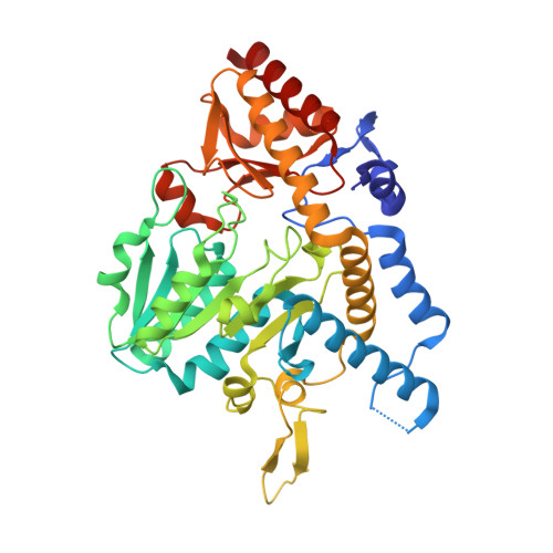
7RRN, 9D2D - PubMed Abstract:
Under stress conditions, iron-sulfur cluster biogenesis in Escherichia coli is initiated by the cysteine desulfurase, SufS, via the SUF pathway. SufS is a type II cysteine desulfurase that catalyzes the PLP-dependent breakage of an l-cysteine C-S bond to generate l-alanine and a covalent active site persulfide. The cysteine desulfurase activity of SufS is activated by SufE, which accepts the covalent persulfide from SufS to regenerate the active site. Based on analysis of the SufS/SufE structure, it was hypothesized that two conserved arginine residues in the SufS active site, R56 and R359, could be important for persulfide transfer from SufS to SufE by regulating the positioning of the α3-α4 loop on SufS. To investigate this hypothesis, site-directed mutagenesis was used to obtain R56A/K and R359A/K SufS variants. Alanine substitution at either position caused defects to SufE-dependent SufS activity, with more conservative lysine substitutions resulting in varying levels of rescued activity. Fluorescence polarization binding assays showed that the loss of SufS activity was not due to a defect in forming the SufS/SufE complex. Surprisingly, the R359A substitution resulted in a 10-fold improvement in the K D value for complex formation. The structure of R359A SufS explains this result as it exhibits a conformational change in the α3-α4 loop allowing SufE better access to the SufS active site. Taken together, the kinetic, binding, and structural data support a mechanism where R359 plays a role in linking SufS catalysis with modulation of the α3-α4 loop to promote a close-approach interaction of SufS and SufE conducive to persulfide transfer.
- Department of Chemistry & Biochemistry, The University of Alabama, Tuscaloosa, Alabama 35487, United States.
Organizational Affiliation: