Structural Characterization of the Human Cytosolic Malate Dehydrogenase I.
McCue, W.M., Finzel, B.C.(2022) ACS Omega 7: 207-214
- PubMed: 35036692 Search on PubMedSearch on PubMed Central
- DOI: https://doi.org/10.1021/acsomega.1c04385
- Primary Citation Related Structures:
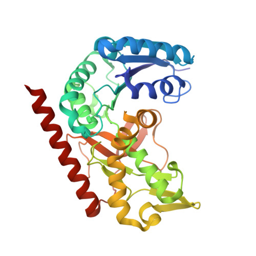
7RM9 - PubMed Abstract:
The first crystal structure of the human cytosolic malate dehydrogenase I (MDH1) is described. Structure determination at a high resolution (1.65 Å) followed production, isolation, and purification of human MDH1 using a bacterial expression system. The structure is a binary complex of MDH1 with only a bound malonate molecule in the substrate binding site. Comparisons of this structure with malate dehydrogenase enzymes from other species confirm that the human enzyme adopts similar secondary, tertiary, and quaternary structures and that the enzyme retains a similar conformation even when nicotinamide adenine dinucleotide (NAD + ) is not bound. A comparison to the highly homologous porcine ( sus scrofa ) MDH1 ternary structures leads to the conclusion that only small conformational differences are needed to accommodate binding by NAD + or other NAD + mimetics. Conformational differences observed in the second subunit show that the NAD + binding elements are nevertheless quite flexible. Comparison of h MDH1 to the human mitochondrial malate dehydrogenase ( h MDH2) reveals some key differences in the α7-α8 loop, which lies directly beneath the substrate binding pocket. These differences might be exploited in the structure-assisted design of selective small molecule inhibitors of h MDH1, an emerging target for the development of anticancer therapeutics.
- Department of Medicinal Chemistry, University of Minnesota, 308 Harvard Street SE, Minneapolis, Minnesota 55455, United States.
Organizational Affiliation: