A structural model of the human plasminogen and Aspergillus fumigatus enolase complex.
Nguyen, S., Jovcevski, B., Truong, J.Q., Pukala, T.L., Bruning, J.B.(2022) Proteins 90: 1509-1520
- PubMed: 35247004 Search on PubMed
- DOI: https://doi.org/10.1002/prot.26331
- Primary Citation Related Structures:
7RHV, 7RHW, 7RI0 - PubMed Abstract:
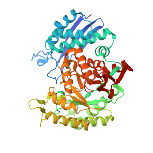
The metabolic enzyme, enolase, plays a crucial role in the cytoplasm where it maintains cellular energy production within the process of glycolysis. The main role of enolase in glycolysis is to convert 2-phosphoglycerate to phosphoenolpyruvate; however, enolase can fulfill roles that deviate from this function. In pathogenic bacteria and fungi, enolase is also located on the cell surface where it functions as a virulence factor. Surface-expressed enolase is a receptor for human plasma proteins, including plasminogen, and this interaction facilitates nutrient acquisition and tissue invasion. A novel approach to developing antifungal drugs is to inhibit the formation of this complex. To better understand the structure of enolase and the interactions that may govern complex formation, we have solved the first X-ray crystal structure of enolase from Aspergillus fumigatus (2.0 Å) and have shown that it preferentially adopts a dimeric quaternary structure using native mass spectrometry. Two additional X-ray crystal structures of A. fumigatus enolase bound to the endogenous substrate 2-phosphoglycerate and product phosphoenolpyruvate were determined and kinetic characterization was carried out to better understand the details of its canonical function. From these data, we have produced a model of the A. fumigatus enolase and human plasminogen complex to provide structural insights into the mechanisms of virulence and aid future development of small molecules or peptidomimetics for antifungal drug design.
- Institute of Photonics and Advanced Sensing (IPAS), School of Biological Sciences, The University of Adelaide, Adelaide, South Australia, Australia.
Organizational Affiliation: