7RDW | pdb_00007rdw

Crystal Structure of FH1 Fab bound to HXb2 HIV-1 gp120 core


Experimental Data Snapshot

  • Method: X-RAY DIFFRACTION
  • Resolution: 3.55 Å
  • R-Value Free: 
    0.295 (Depositor), 0.294 (DCC) 
  • R-Value Work: 
    0.260 (Depositor), 0.262 (DCC) 
  • R-Value Observed: 
    0.262 (Depositor) 

Starting Model: experimental
View more details

wwPDB Validation 3D Report Full Report

Validation slider image for 7RDW

Ligand Structure Quality Assessment 


This is version 1.2 of the entry. See complete history

Literature

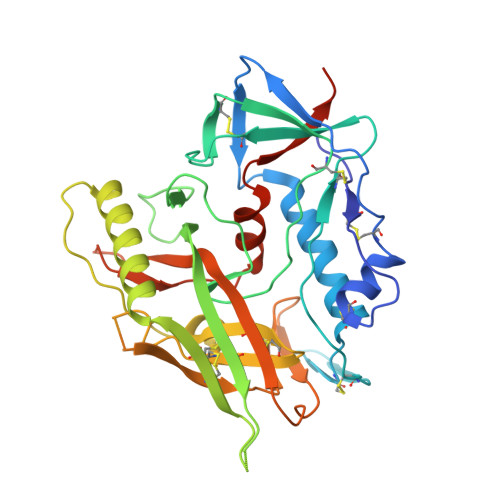
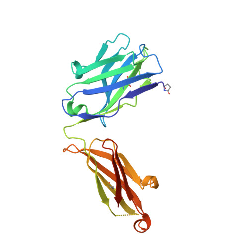
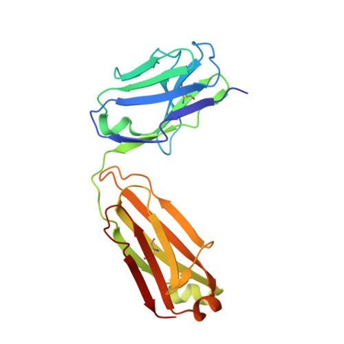
Characterization of a vaccine-elicited human antibody with sequence homology to VRC01-class antibodies that binds the C1C2 gp120 domain.

Gray, M.D.Feng, J.Weidle, C.E.Cohen, K.W.Ballweber-Fleming, L.MacCamy, A.J.Huynh, C.N.Trichka, J.J.Montefiori, D.Ferrari, G.Pancera, M.McElrath, M.J.Stamatatos, L.

(2022) Sci Adv 8: eabm3948-eabm3948

  • DOI: https://doi.org/10.1126/sciadv.abm3948
  • Primary Citation Related Structures: 
    7RDW

  • PubMed Abstract: 

    Broadly HIV-1-neutralizing VRC01-class antibodies bind the CD4-binding site of Env and contain V H 1-2*02-derived heavy chains paired with light chains expressing five-amino acid-long CDRL3s. Their unmutated germline forms do not recognize HIV-1 Env, and their lack of elicitation in human clinical trials could be due to the absence of activation of the corresponding naïve B cells by the vaccine immunogens. To address this point, we examined Env-specific B cell receptor sequences from participants in the HVTN 100 clinical trial. Of all the sequences analyzed, only one displayed homology to VRC01-class antibodies, but the corresponding antibody (FH1) recognized the C1C2 gp120 domain. For FH1 to switch epitope recognition to the CD4-binding site, alterations in the CDRH3 and CDRL3 were necessary. Only germ line-targeting Env immunogens efficiently activated VRC01 B cells, even in the presence of FH1 B cells. Our findings support the use of these immunogens to activate VRC01 B cells in humans.


  • Organizational Affiliation
    • Vaccine and Infectious Disease Division, Fred Hutchinson Cancer Research Center, Seattle, WA 98109, USA.

Macromolecule Content 

  • Total Structure Weight: 365.29 kDa 
  • Atom Count: 21,960 
  • Modeled Residue Count: 2,973 
  • Deposited Residue Count: 3,236 
  • Unique protein chains: 4

Macromolecules

Find similar proteins by:|  3D Structure
Entity ID: 1
MoleculeChains  Sequence LengthOrganismDetailsImage
Glycoprotein 120A [auth C],
B [auth D],
G [auth M],
H [auth N]
373Human immunodeficiency virus 1Mutation(s): 0 
UniProt
Find proteins for P04578 (Human immunodeficiency virus type 1 group M subtype B (isolate HXB2))
Explore P04578 
Go to UniProtKB:  P04578
Entity Groups
Sequence Clusters30% Identity50% Identity70% Identity90% Identity95% Identity100% Identity
UniProt GroupP04578
Glycosylation
Glycosylation Sites: 11
Sequence Annotations
Expand
Reference Sequence
Find similar proteins by:|  3D Structure
Entity ID: 2
MoleculeChains  Sequence LengthOrganismDetailsImage
FH1 Fab Heavy ChainC [auth G],
K [auth U]
225Homo sapiensMutation(s): 0 
Find similar proteins by:|  3D Structure
Entity ID: 3
MoleculeChains  Sequence LengthOrganismDetailsImage
FH1 Fab Heavy ChainD [auth H],
I [auth Q]
225Homo sapiensMutation(s): 0 
Find similar proteins by:|  3D Structure
Entity ID: 4
MoleculeChains  Sequence LengthOrganismDetailsImage
FH1 Fab Light ChainE [auth I],
F [auth L],
J [auth R],
L [auth V]
211Homo sapiensMutation(s): 0 

Oligosaccharides

Help  
Entity ID: 5
MoleculeChains Length2D Diagram GlycosylationD Interactions
beta-D-mannopyranose-(1-4)-2-acetamido-2-deoxy-beta-D-glucopyranose-(1-4)-2-acetamido-2-deoxy-beta-D-glucopyranoseM [auth O],
N [auth c],
O [auth m],
P [auth y]
3N-Glycosylation
Glycosylation Resources
GlyTouCan: G15407YE
GlyCosmos: G15407YE
GlyGen: G15407YE

Small Molecules

Ligands 4 Unique
IDChains Name / Formula / InChI Key2D Diagram3D Interactions
NAG

Query on NAG


Download:Ideal Coordinates CCD File
AB [auth N]
BB [auth N]
CA [auth D]
CB [auth N]
DA [auth D]
AB [auth N],
BB [auth N],
CA [auth D],
CB [auth N],
DA [auth D],
DB [auth N],
EA [auth D],
FA [auth D],
GA [auth D],
NA [auth M],
OA [auth M],
PA [auth M],
Q [auth C],
QA [auth M],
R [auth C],
RA [auth M],
S [auth C],
SA [auth M],
T [auth C],
U [auth C],
UA [auth N],
V [auth C],
VA [auth N],
W [auth C],
WA [auth N],
XA [auth N],
YA [auth N],
ZA [auth N]
2-acetamido-2-deoxy-beta-D-glucopyranose
C8 H15 N O6
OVRNDRQMDRJTHS-FMDGEEDCSA-N
SO4

Query on SO4


Download:Ideal Coordinates CCD File
BA [auth C],
JA [auth D],
TA [auth M]
SULFATE ION
O4 S
QAOWNCQODCNURD-UHFFFAOYSA-L
EDO

Query on EDO


Download:Ideal Coordinates CCD File
AA [auth C]
EB [auth N]
IA [auth D]
KA [auth H]
LA [auth H]
AA [auth C],
EB [auth N],
IA [auth D],
KA [auth H],
LA [auth H],
Z [auth C]
1,2-ETHANEDIOL
C2 H6 O2
LYCAIKOWRPUZTN-UHFFFAOYSA-N
NH4

Query on NH4


Download:Ideal Coordinates CCD File
HA [auth D],
MA [auth I],
X [auth C],
Y [auth C]
AMMONIUM ION
H4 N
QGZKDVFQNNGYKY-UHFFFAOYSA-O
Modified Residues  1 Unique
IDChains TypeFormula2D DiagramParent
PCA
Query on PCA
C [auth G],
K [auth U]
L-PEPTIDE LINKINGC5 H7 N O3GLN

Experimental Data & Validation

Experimental Data

  • Method: X-RAY DIFFRACTION
  • Resolution: 3.55 Å
  • R-Value Free:  0.295 (Depositor), 0.294 (DCC) 
  • R-Value Work:  0.260 (Depositor), 0.262 (DCC) 
  • R-Value Observed: 0.262 (Depositor) 
Space Group: P 1
Unit Cell:
Length ( Å )Angle ( ˚ )
a = 103.361α = 67.566
b = 106.266β = 76.674
c = 113.219γ = 60.987
Software Package:
Software NamePurpose
PHENIXrefinement
HKL-2000data reduction
SCALEPACKdata scaling
Cootmodel building
PHASERphasing

Structure Validation

View Full Validation Report


Ligand Structure Quality Assessment 


Entry History 

& Funding Information

Deposition Data


Funding OrganizationLocationGrant Number
National Institutes of Health/National Institute Of Allergy and Infectious Diseases (NIH/NIAID)United States--

Revision History  (Full details and data files)

  • Version 1.0: 2022-05-25
    Type: Initial release
  • Version 1.1: 2023-10-18
    Changes: Data collection, Refinement description
  • Version 1.2: 2024-10-23
    Changes: Structure summary