Conformational equilibria in allosteric control of Hsp70 chaperones.
Wang, W., Liu, Q., Liu, Q., Hendrickson, W.A.(2021) Mol Cell 81: 3919-3933.e7
- PubMed: 34453889 Search on PubMedSearch on PubMed Central
- DOI: https://doi.org/10.1016/j.molcel.2021.07.039
- Primary Citation Related Structures:
7KO2, 7KRT, 7KRU, 7KRV, 7KRW, 7N46, 7RAX - PubMed Abstract:
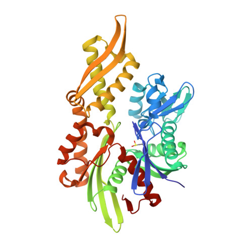
Heat-shock proteins of 70 kDa (Hsp70s) are vital for all life and are notably important in protein folding. Hsp70s use ATP binding and hydrolysis at a nucleotide-binding domain (NBD) to control the binding and release of client polypeptides at a substrate-binding domain (SBD); however, the mechanistic basis for this allostery has been elusive. Here, we first characterize biochemical properties of selected domain-interface mutants in bacterial Hsp70 DnaK. We then develop a theoretical model for allosteric equilibria among Hsp70 conformational states to explain the observations: a restraining state, Hsp70 R -ATP, restricts ATP hydrolysis and binds peptides poorly, whereas a stimulating state, Hsp70 S -ATP, hydrolyzes ATP rapidly and has high intrinsic substrate affinity but rapid binding kinetics. We support this model for allosteric regulation with DnaK structures obtained in the postulated stimulating state S with biochemical tests of the S-state interface and with improved peptide-binding-site definition in an R-state structure.
- Department of Biochemistry and Molecular Biophysics, Columbia University, New York, NY 10032, USA; Department of Biological Sciences, Columbia University, New York, NY 10027, USA.
Organizational Affiliation: