7R07

Abortive infection DNA polymerase AbiK from Lactococcus lactis


Experimental Data Snapshot

  • Method: X-RAY DIFFRACTION
  • Resolution: 3.10 Å
  • R-Value Free: 0.232 
  • R-Value Work: 0.189 
  • R-Value Observed: 0.190 

wwPDB Validation   3D Report Full Report


This is version 1.3 of the entry. See complete history


Literature

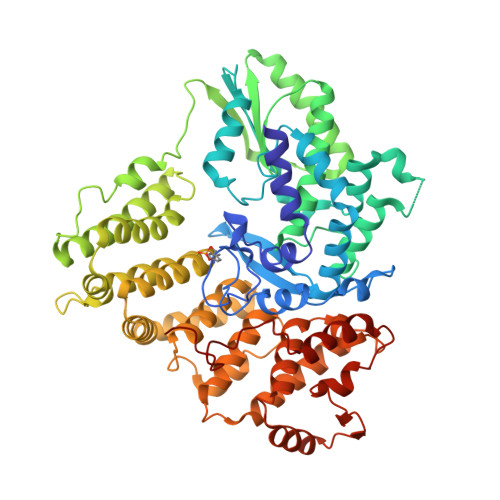
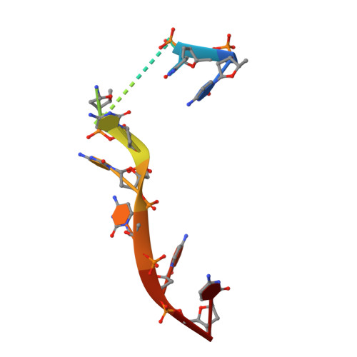
Mechanism of protein-primed template-independent DNA synthesis by Abi polymerases.

Figiel, M.Gapinska, M.Czarnocki-Cieciura, M.Zajko, W.Sroka, M.Skowronek, K.Nowotny, M.

(2022) Nucleic Acids Res 50: 10026-10040

  • DOI: https://doi.org/10.1093/nar/gkac772
  • Primary Citation of Related Structures:  
    7R06, 7R07, 7R08, 7Z0Z

  • PubMed Abstract: 

    Abortive infection (Abi) is a bacterial antiphage defense strategy involving suicide of the infected cell. Some Abi pathways involve polymerases that are related to reverse transcriptases. They are unique in the way they combine the ability to synthesize DNA in a template-independent manner with protein priming. Here, we report crystal and cryo-electron microscopy structures of two Abi polymerases: AbiK and Abi-P2. Both proteins adopt a bilobal structure with an RT-like domain that comprises palm and fingers subdomains and a unique helical domain. AbiK and Abi-P2 adopt a hexameric and trimeric configuration, respectively, which is unprecedented for reverse transcriptases. Biochemical experiments showed that the formation of these oligomers is required for the DNA polymerization activity. The structure of the AbiK-DNA covalent adduct visualized interactions between the 3' end of DNA and the active site and covalent attachment of the 5' end of DNA to a tyrosine residue used for protein priming. Our data reveal a structural basis of the mechanism of highly unusual template-independent protein-priming polymerases.


  • Organizational Affiliation

    Laboratory of Protein Structure, International Institute of Molecular and Cell Biology, Warsaw, Poland.


Macromolecules

Find similar proteins by:  (by identity cutoff)  |  3D Structure
Entity ID: 1
MoleculeChains Sequence LengthOrganismDetailsImage
AbiK
A, B, C, D, E
A, B, C, D, E, F
601Lactococcus lactisMutation(s): 0 
Gene Names: abiK
UniProt
Find proteins for Q48614 (Lactococcus lactis)
Explore Q48614 
Go to UniProtKB:  Q48614
Entity Groups  
Sequence Clusters30% Identity50% Identity70% Identity90% Identity95% Identity100% Identity
UniProt GroupQ48614
Sequence Annotations
Expand
  • Reference Sequence

Find similar nucleic acids by:  Sequence   |   3D Structure  

Entity ID: 2
MoleculeChains LengthOrganismImage
DNA (5'-D(*CP*CP*CP*CP*CP*CP*CP*CP*CP*CP*C)-3')
G, H, I, J, K
G, H, I, J, K, L
11Escherichia coli BL21
Sequence Annotations
Expand
  • Reference Sequence
Experimental Data & Validation

Experimental Data

  • Method: X-RAY DIFFRACTION
  • Resolution: 3.10 Å
  • R-Value Free: 0.232 
  • R-Value Work: 0.189 
  • R-Value Observed: 0.190 
  • Space Group: P 62 2 2
Unit Cell:
Length ( Å )Angle ( ˚ )
a = 200.99α = 90
b = 200.99β = 90
c = 570.72γ = 120
Software Package:
Software NamePurpose
PHENIXrefinement
XDSdata reduction
XSCALEdata scaling
PHASERphasing

Structure Validation

View Full Validation Report


Entry History & Funding Information

Deposition Data


Funding OrganizationLocationGrant Number
Foundation for Polish SciencePolandPOIR.04.04.00-00-20E7/16-00
European Regional Development FundEuropean UnionPOIG.02.02.00-14-024/08-00

Revision History  (Full details and data files)

  • Version 1.0: 2022-09-07
    Type: Initial release
  • Version 1.1: 2022-09-28
    Changes: Database references
  • Version 1.2: 2022-10-05
    Changes: Database references
  • Version 1.3: 2024-05-01
    Changes: Data collection, Refinement description