Pseudouridine-Modifying Enzymes SapB and SapH Control Entry into the Pseudouridimycin Biosynthetic Pathway.
Artukka, E., Schnell, R., Palmu, K., Rosenqvist, P., Szodorai, E., Niemi, J., Virta, P., Schneider, G., Metsa-Ketela, M.(2023) ACS Chem Biol 18: 794-802
- PubMed: 37005433 Search on PubMedSearch on PubMed Central
- DOI: https://doi.org/10.1021/acschembio.2c00826
- Primary Citation Related Structures:
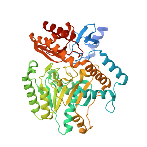
7QZJ - PubMed Abstract:
Pseudouridimycin is a microbial C -nucleoside natural product that specifically inhibits bacterial RNA polymerases by binding to the active site and competing with uridine triphosphate for the nucleoside triphosphate (NTP) addition site. Pseudouridimycin consists of 5'-aminopseudouridine and formamidinylated, N-hydroxylated Gly-Gln dipeptide moieties to allow Watson-Crick base pairing and to mimic protein-ligand interactions of the triphosphates of NTP, respectively. The metabolic pathway of pseudouridimycin has been studied in Streptomyces species, but no biosynthetic steps have been characterized biochemically. Here, we show that the flavin-dependent oxidase SapB functions as a gate-keeper enzyme selecting pseudouridine ( K M = 34 μM) over uridine ( K M = 901 μM) in the formation of pseudouridine aldehyde. The pyridoxal phosphate (PLP)-dependent SapH catalyzes transamination, resulting in 5'-aminopseudouridine with a preference for arginine, methionine, or phenylalanine as cosubstrates as amino group donors. The binary structure of SapH in complex with pyridoxamine-5'-phosphate and site-directed mutagenesis identified Lys289 and Trp32 as key residues for catalysis and substrate binding, respectively. The related C -nucleoside oxazinomycin was accepted as a substrate by SapB with moderate affinity ( K M = 181 μM) and was further converted by SapH, which opens possibilities for metabolic engineering to generate hybrid C -nucleoside pseudouridimycin analogues in Streptomyces .
- Department of Life Technologies, University of Turku, FIN-20014 Turku, Finland.
Organizational Affiliation: