7QUR

SARS-CoV-2 Spike with ethylbenzamide-tri-iodo Siallyllactose, C3 symmetry


Experimental Data Snapshot

  • Method: ELECTRON MICROSCOPY
  • Resolution: 2.27 Å
  • Aggregation State: PARTICLE 
  • Reconstruction Method: SINGLE PARTICLE 

wwPDB Validation   3D Report Full Report


This is version 1.3 of the entry. See complete history


Literature

Pathogen-sugar interactions revealed by universal saturation transfer analysis.

Buchanan, C.J.Gaunt, B.Harrison, P.J.Yang, Y.Liu, J.Khan, A.Giltrap, A.M.Le Bas, A.Ward, P.N.Gupta, K.Dumoux, M.Tan, T.K.Schimaski, L.Daga, S.Picchiotti, N.Baldassarri, M.Benetti, E.Fallerini, C.Fava, F.Giliberti, A.Koukos, P.I.Davy, M.J.Lakshminarayanan, A.Xue, X.Papadakis, G.Deimel, L.P.Casablancas-Antras, V.Claridge, T.D.W.Bonvin, A.M.J.J.Sattentau, Q.J.Furini, S.Gori, M.Huo, J.Owens, R.J.Schaffitzel, C.Berger, I.Renieri, A.Naismith, J.H.Baldwin, A.J.Davis, B.G.

(2022) Science 377: eabm3125-eabm3125

  • DOI: https://doi.org/10.1126/science.abm3125
  • Primary Citation of Related Structures:  
    7QUR, 7QUS

  • PubMed Abstract: 

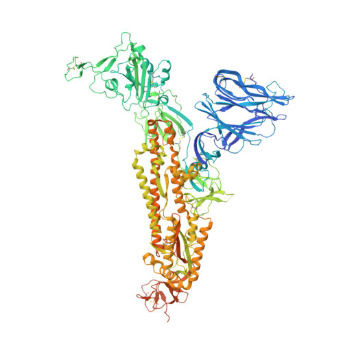
    Many pathogens exploit host cell-surface glycans. However, precise analyses of glycan ligands binding with heavily modified pathogen proteins can be confounded by overlapping sugar signals and/or compounded with known experimental constraints. Universal saturation transfer analysis (uSTA) builds on existing nuclear magnetic resonance spectroscopy to provide an automated workflow for quantitating protein-ligand interactions. uSTA reveals that early-pandemic, B-origin-lineage severe acute respiratory syndrome coronavirus 2 (SARS-CoV-2) spike trimer binds sialoside sugars in an "end-on" manner. uSTA-guided modeling and a high-resolution cryo-electron microscopy structure implicate the spike N-terminal domain (NTD) and confirm end-on binding. This finding rationalizes the effect of NTD mutations that abolish sugar binding in SARS-CoV-2 variants of concern. Together with genetic variance analyses in early pandemic patient cohorts, this binding implicates a sialylated polylactosamine motif found on tetraantennary N-linked glycoproteins deep in the human lung as potentially relevant to virulence and/or zoonosis.


  • Organizational Affiliation

    Rosalind Franklin Institute, Harwell Science and Innovation Campus, Oxford OX11 0FA, UK.


Macromolecules
Find similar proteins by:  (by identity cutoff)  |  3D Structure
Entity ID: 1
MoleculeChains Sequence LengthOrganismDetailsImage
Spike glycoprotein,Fibritin
A, B, C
1,259Severe acute respiratory syndrome coronavirus 2Tequatrovirus T4
This entity is chimeric
Mutation(s): 1 
Gene Names: S2wac
UniProt
Find proteins for P0DTC2 (Severe acute respiratory syndrome coronavirus 2)
Explore P0DTC2 
Go to UniProtKB:  P0DTC2
Entity Groups  
Sequence Clusters30% Identity50% Identity70% Identity90% Identity95% Identity100% Identity
UniProt GroupP0DTC2
Sequence Annotations
Expand
  • Reference Sequence
Oligosaccharides

Help

Entity ID: 2
MoleculeChains Length2D Diagram Glycosylation3D Interactions
2-acetamido-2-deoxy-beta-D-glucopyranose-(1-4)-2-acetamido-2-deoxy-beta-D-glucopyranose
D, E, F, H, I
D, E, F, H, I, J, L, M, N
2N-Glycosylation
Glycosylation Resources
GlyTouCan:  G42666HT
GlyCosmos:  G42666HT
GlyGen:  G42666HT
Entity ID: 3
MoleculeChains Length2D Diagram Glycosylation3D Interactions
alpha-L-fucopyranose-(1-3)-[2-acetamido-2-deoxy-beta-D-glucopyranose-(1-4)][alpha-L-fucopyranose-(1-6)]2-acetamido-2-deoxy-beta-D-glucopyranose
G, K, O
4N-Glycosylation
Glycosylation Resources
GlyTouCan:  G63564LA
GlyCosmos:  G63564LA
Small Molecules
Ligands 4 Unique
IDChains Name / Formula / InChI Key2D Diagram3D Interactions
GE9 (Subject of Investigation/LOI)
Query on GE9

Download Ideal Coordinates CCD File 
BA [auth A],
GB [auth C],
QA [auth B]
2,3,5-tris(iodanyl)benzamide
C7 H4 I3 N O
OVJSATAEGZSMIJ-UHFFFAOYSA-N
SIA (Subject of Investigation/LOI)
Query on SIA

Download Ideal Coordinates CCD File 
CA [auth A],
HB [auth C],
RA [auth B]
N-acetyl-alpha-neuraminic acid
C11 H19 N O9
SQVRNKJHWKZAKO-YRMXFSIDSA-N
EIC
Query on EIC

Download Ideal Coordinates CCD File 
DA [auth A],
SA [auth B],
TA [auth C]
LINOLEIC ACID
C18 H32 O2
OYHQOLUKZRVURQ-HZJYTTRNSA-N
NAG
Query on NAG

Download Ideal Coordinates CCD File 
AA [auth A]
AB [auth C]
BB [auth C]
CB [auth C]
DB [auth C]
AA [auth A],
AB [auth C],
BB [auth C],
CB [auth C],
DB [auth C],
EA [auth B],
EB [auth C],
FA [auth B],
FB [auth C],
GA [auth B],
HA [auth B],
IA [auth B],
JA [auth B],
KA [auth B],
LA [auth B],
MA [auth B],
NA [auth B],
OA [auth B],
P [auth A],
PA [auth B],
Q [auth A],
R [auth A],
S [auth A],
T [auth A],
U [auth A],
UA [auth C],
V [auth A],
VA [auth C],
W [auth A],
WA [auth C],
X [auth A],
XA [auth C],
Y [auth A],
YA [auth C],
Z [auth A],
ZA [auth C]
2-acetamido-2-deoxy-beta-D-glucopyranose
C8 H15 N O6
OVRNDRQMDRJTHS-FMDGEEDCSA-N
Experimental Data & Validation

Experimental Data

  • Method: ELECTRON MICROSCOPY
  • Resolution: 2.27 Å
  • Aggregation State: PARTICLE 
  • Reconstruction Method: SINGLE PARTICLE 

Structure Validation

View Full Validation Report


Entry History & Funding Information

Deposition Data


Funding OrganizationLocationGrant Number
Other government--

Revision History  (Full details and data files)

  • Version 1.0: 2022-06-01
    Type: Initial release
  • Version 1.1: 2022-06-15
    Changes: Database references, Source and taxonomy, Structure summary
  • Version 1.2: 2022-07-06
    Changes: Database references
  • Version 1.3: 2022-08-03
    Changes: Database references