7QPN

Crystal Structure of Phosphatidylinositol 5-Phosphate 4-Kinase (PI5P4K2C) bound to an allosteric inhibitor and AMP-PNP


Experimental Data Snapshot

  • Method: X-RAY DIFFRACTION
  • Resolution: 1.95 Å
  • R-Value Free: 0.262 
  • R-Value Work: 0.192 
  • R-Value Observed: 0.195 

wwPDB Validation   3D Report Full Report


Ligand Structure Quality Assessment 


This is version 1.3 of the entry. See complete history


Literature

Development of Selective Phosphatidylinositol 5-Phosphate 4-Kinase gamma Inhibitors with a Non-ATP-competitive, Allosteric Binding Mode.

Boffey, H.K.Rooney, T.P.C.Willems, H.M.G.Edwards, S.Green, C.Howard, T.Ogg, D.Romero, T.Scott, D.E.Winpenny, D.Duce, J.Skidmore, J.Clarke, J.H.Andrews, S.P.

(2022) J Med Chem 65: 3359-3370

  • DOI: https://doi.org/10.1021/acs.jmedchem.1c01819
  • Primary Citation of Related Structures:  
    7QIE, 7QPN

  • PubMed Abstract: 

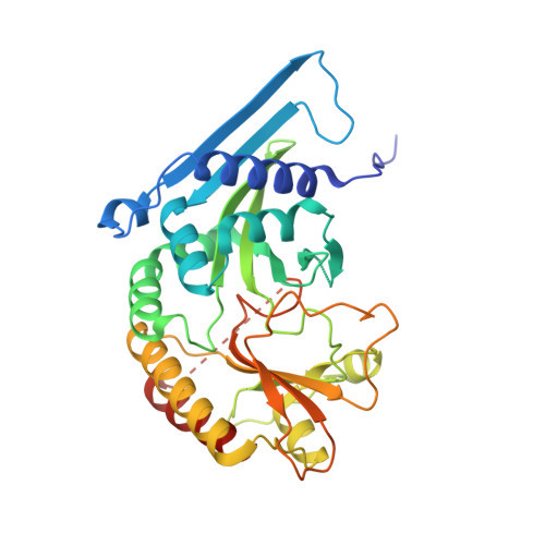
    Phosphatidylinositol 5-phosphate 4-kinases (PI5P4Ks) are emerging as attractive therapeutic targets in diseases, such as cancer, immunological disorders, and neurodegeneration, owing to their central role in regulating cell signaling pathways that are either dysfunctional or can be modulated to promote cell survival. Different modes of binding may enhance inhibitor selectivity and reduce off-target effects in cells. Here, we describe efforts to improve the physicochemical properties of the selective PI5P4Kγ inhibitor, NIH-12848 ( 1 ). These improvements enabled the demonstration that this chemotype engages PI5P4Kγ in intact cells and that compounds from this series do not inhibit PI5P4Kα or PI5P4Kβ. Furthermore, the first X-ray structure of PI5P4Kγ bound to an inhibitor has been determined with this chemotype, confirming an allosteric binding mode. An exemplar from this chemical series adopted two distinct modes of inhibition, including through binding to a putative lipid interaction site which is 18 Å from the ATP pocket.


  • Organizational Affiliation

    The ALBORADA Drug Discovery Institute, University of Cambridge, Island Research Building, Cambridge Biomedical Campus, Hills Road, Cambridge CB2 0AH, U.K.


Macromolecules
Find similar proteins by:  (by identity cutoff)  |  3D Structure
Entity ID: 1
MoleculeChains Sequence LengthOrganismDetailsImage
Phosphatidylinositol 5-Phosphate 4-Kinase (PI5P4K2C)
A, B
348Homo sapiensMutation(s): 0 
UniProt & NIH Common Fund Data Resources
Find proteins for Q8TBX8 (Homo sapiens)
Explore Q8TBX8 
Go to UniProtKB:  Q8TBX8
PHAROS:  Q8TBX8
GTEx:  ENSG00000166908 
Entity Groups  
Sequence Clusters30% Identity50% Identity70% Identity90% Identity95% Identity100% Identity
UniProt GroupQ8TBX8
Sequence Annotations
Expand
  • Reference Sequence
Experimental Data & Validation

Experimental Data

  • Method: X-RAY DIFFRACTION
  • Resolution: 1.95 Å
  • R-Value Free: 0.262 
  • R-Value Work: 0.192 
  • R-Value Observed: 0.195 
  • Space Group: P 1 21 1
Unit Cell:
Length ( Å )Angle ( ˚ )
a = 47.921α = 90
b = 65.625β = 93.16
c = 117.12γ = 90
Software Package:
Software NamePurpose
REFMACrefinement
PDB_EXTRACTdata extraction
XDSdata reduction
Aimlessdata scaling
PHASERphasing

Structure Validation

View Full Validation Report


Ligand Structure Quality Assessment 


Entry History & Funding Information

Deposition Data


Funding OrganizationLocationGrant Number
Alzheimers Research UK (ARUK)United KingdomARUK-2015DDI-CAM

Revision History  (Full details and data files)

  • Version 1.0: 2022-05-18
    Type: Initial release
  • Version 1.1: 2022-08-10
    Changes: Database references
  • Version 1.2: 2022-08-17
    Changes: Database references
  • Version 1.3: 2024-01-31
    Changes: Data collection, Refinement description