Medical contrast agents as promising tools for biomacromolecular SAXS experiments.
Gabel, F., Engilberge, S., Schmitt, E., Thureau, A., Mechulam, Y., Perez, J., Girard, E.(2022) Acta Crystallogr D Struct Biol 78: 1120-1130
- PubMed: 36048152 Search on PubMedSearch on PubMed Central
- DOI: https://doi.org/10.1107/S2059798322007392
- Primary Citation Related Structures:
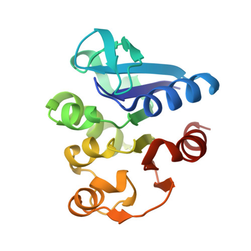
7QO8 - PubMed Abstract:
Small-angle X-ray scattering (SAXS) has become an indispensable tool in structural biology, complementing atomic-resolution techniques. It is sensitive to the electron-density difference between solubilized biomacromolecules and the buffer, and provides information on molecular masses, particle dimensions and interactions, low-resolution conformations and pair distance-distribution functions. When SAXS data are recorded at multiple contrasts, i.e. at different solvent electron densities, it is possible to probe, in addition to their overall shape, the internal electron-density profile of biomacromolecular assemblies. Unfortunately, contrast-variation SAXS has been limited by the range of solvent electron densities attainable using conventional co-solutes (for example sugars, glycerol and salt) and by the fact that some biological systems are destabilized in their presence. Here, SAXS contrast data from an oligomeric protein and a protein-RNA complex are presented in the presence of iohexol and Gd-HPDO3A, two electron-rich molecules that are used in biomedical imaging and that belong to the families of iodinated and lanthanide-based complexes, respectively. Moderate concentrations of both molecules allowed solvent electron densities matching those of proteins to be attained. While iohexol yielded higher solvent electron densities (per mole), it interacted specifically with the oligomeric protein and precipitated the protein-RNA complex. Gd-HPDO3A, while less efficient (per mole), did not disrupt the structural integrity of either system, and atomic models could be compared with the SAXS data. Due to their elevated solubility and electron density, their chemical inertness, as well as the possibility of altering their physico-chemical properties, lanthanide-based complexes represent a class of molecules with promising potential for contrast-variation SAXS experiments on diverse biomacromolecular systems.
- IBS, CEA, CNRS, UGA, 71 Avenue des Martyrs, 38000 Grenoble, France.
Organizational Affiliation: