7QNV | pdb_00007qnv

human carbonic anhydrase II bound to 3-methylbenzoselenoate


Experimental Data Snapshot

  • Method: X-RAY DIFFRACTION
  • Resolution: 1.28 Å
  • R-Value Free: 
    0.184 (Depositor), 0.184 (DCC) 
  • R-Value Work: 
    0.164 (Depositor), 0.163 (DCC) 

Starting Model: experimental
View more details

wwPDB Validation 3D Report Full Report

Validation slider image for 7QNV

Ligand Structure Quality Assessment 


This is version 1.2 of the entry. See complete history

Literature

Benzoselenoates: A novel class of carbonic anhydrase inhibitors.

Tanini, D.Capperucci, A.Locuoco, M.Ferraroni, M.Costantino, G.Angeli, A.Supuran, C.T.

(2022) Bioorg Chem 122: 105751-105751

  • DOI: https://doi.org/10.1016/j.bioorg.2022.105751
  • Primary Citation Related Structures: 
    7QNV, 7QOB

  • PubMed Abstract: 

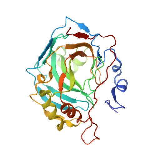
    A series of benzoselenoates has been prepared and their inhibitory properties against the most relevant human Carbonic Anhydrases (CAs) isoforms, among which hCA I, II, IV, VII, IX, and XII were investigated. These inhibitors were designed considering the carboxylates and mono-/dithiocarbamates as lead and led to the observation that the COSe - is a new zinc-binding group (ZBG) for metalloenzymes possessing zinc ions at their active site. The substitution pattern on aromatic ring of the benzoselenoates is the crucial structural element influencing selectivity towards various isoforms. We elucidated the binding mode of benzoselenoates to hCA I and hCA II by using X-ray crystallography. The negatively charged selenium atom from the new ZBG was observed coordinated to the zinc ion from the CA active site at a distance of 2.30-2.40 Å from it. Overall, these data might be useful for the development of new inhibitors with higher selectivity and efficacy for various hCAs.


  • Organizational Affiliation
    • University of Florence, Department of Chemistry "Ugo Schiff", Via della Lastruccia 3-13, I-50019, Sesto Fiorentino, Italy.

Macromolecule Content 

  • Total Structure Weight: 29.84 kDa 
  • Atom Count: 2,354 
  • Modeled Residue Count: 258 
  • Deposited Residue Count: 260 
  • Unique protein chains: 1

Macromolecules

Find similar proteins by:|  3D Structure
Entity ID: 1
MoleculeChains  Sequence LengthOrganismDetailsImage
Carbonic anhydrase 2A [auth AAA]260Homo sapiensMutation(s): 0 
Gene Names: CA2
EC: 4.2.1.1 (PDB Primary Data), 4.2.1.69 (UniProt)
UniProt & NIH Common Fund Data Resources
Find proteins for P00918 (Homo sapiens)
Explore P00918 
Go to UniProtKB:  P00918
PHAROS:  P00918
GTEx:  ENSG00000104267 
Entity Groups
Sequence Clusters30% Identity50% Identity70% Identity90% Identity95% Identity100% Identity
UniProt GroupP00918
Sequence Annotations
Expand
Reference Sequence

Experimental Data & Validation

Experimental Data

  • Method: X-RAY DIFFRACTION
  • Resolution: 1.28 Å
  • R-Value Free:  0.184 (Depositor), 0.184 (DCC) 
  • R-Value Work:  0.164 (Depositor), 0.163 (DCC) 
Space Group: P 1 21 1
Unit Cell:
Length ( Å )Angle ( ˚ )
a = 42.43α = 90
b = 41.42β = 104.42
c = 72.13γ = 90
Software Package:
Software NamePurpose
REFMACrefinement
XDSdata reduction
XDSdata scaling
REFMACphasing

Structure Validation

View Full Validation Report


Ligand Structure Quality Assessment 


Entry History 

& Funding Information

Deposition Data


Funding OrganizationLocationGrant Number
Not funded--

Revision History  (Full details and data files)

  • Version 1.0: 2023-01-18
    Type: Initial release
  • Version 1.1: 2023-08-02
    Changes: Database references
  • Version 1.2: 2024-01-31
    Changes: Data collection, Refinement description