7QHL | pdb_00007qhl

Crystal structure of Cyclin-dependent kinase 2/cyclin A in complex with 3,5,7-Substituted pyrazolo[4,3-d]pyrimidine inhibitor 24


Experimental Data Snapshot

  • Method: X-RAY DIFFRACTION
  • Resolution: 1.70 Å
  • R-Value Free: 
    0.224 (Depositor), 0.231 (DCC) 
  • R-Value Work: 
    0.200 (Depositor), 0.209 (DCC) 
  • R-Value Observed: 
    0.200 (Depositor) 

Starting Model: experimental
View more details

wwPDB Validation 3D Report Full Report

Validation slider image for 7QHL

Ligand Structure Quality Assessment 


This is version 1.3 of the entry. See complete history

Literature

3,5,7-Substituted Pyrazolo[4,3- d ]Pyrimidine Inhibitors of Cyclin-Dependent Kinases and Cyclin K Degraders.

Jorda, R.Havlicek, L.Perina, M.Vojackova, V.Pospisil, T.Djukic, S.Skerlova, J.Gruz, J.Renesova, N.Klener, P.Rezacova, P.Strnad, M.Krystof, V.

(2022) J Med Chem 65: 8881-8896

  • DOI: https://doi.org/10.1021/acs.jmedchem.1c02184
  • Primary Citation Related Structures: 
    7QHL

  • PubMed Abstract: 

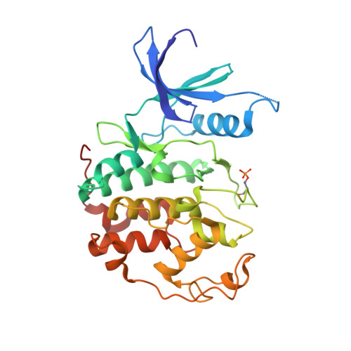
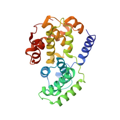
    3,5,7-Trisubstituted pyrazolo[4,3- d ]pyrimidines have been identified as potent inhibitors of cyclin-dependent kinases (CDKs), which are established drug targets. Herein, we describe their further structural modifications leading to novel nanomolar inhibitors with strong antiproliferative activity. We determined the crystal structure of fully active CDK2/A2 with 5-(2-amino-1-ethyl)thio-3-cyclobutyl-7-[4-(pyrazol-1-yl)benzyl]amino-1(2) H -pyrazolo[4,3- d ]pyrimidine ( 24 ) at 1.7 Å resolution, confirming the competitive mode of inhibition. Biochemical and cellular assays in lymphoma cell lines confirmed the expected mechanism of action through dephosphorylation of retinoblastoma protein and RNA polymerase II, leading to induction of apoptosis. Importantly, we also revealed an interesting ability of compound 24 to induce proteasome-dependent degradation of cyclin K both in vitro and in a patient-derived xenograft in vivo. We propose that 24 has a dual mechanism of action, acting as a kinase inhibitor and as a molecular glue inducing an interaction between CDK12 and DDB1 that leads to polyubiquitination of cyclin K and its subsequent degradation.


  • Organizational Affiliation
    • Department of Experimental Biology, Faculty of Science, Palacký University Olomouc, Šlechtitelů 27, 78371 Olomouc, Czech Republic.

Macromolecule Content 

  • Total Structure Weight: 129.58 kDa 
  • Atom Count: 9,960 
  • Modeled Residue Count: 1,086 
  • Deposited Residue Count: 1,114 
  • Unique protein chains: 2

Macromolecules

Find similar proteins by:|  3D Structure
Entity ID: 1
MoleculeChains  Sequence LengthOrganismDetailsImage
Cyclin-dependent kinase 2
A, C
299Homo sapiensMutation(s): 0 
Gene Names: CDK2CDKN2
EC: 2.7.11.22
UniProt & NIH Common Fund Data Resources
Find proteins for P24941 (Homo sapiens)
Explore P24941 
Go to UniProtKB:  P24941
PHAROS:  P24941
GTEx:  ENSG00000123374 
Entity Groups
Sequence Clusters30% Identity50% Identity70% Identity90% Identity95% Identity100% Identity
UniProt GroupP24941
Sequence Annotations
Expand
Reference Sequence
Find similar proteins by:|  3D Structure
Entity ID: 2
MoleculeChains  Sequence LengthOrganismDetailsImage
Cyclin-A2
B, D
258Homo sapiensMutation(s): 0 
Gene Names: CCNA2CCN1CCNA
UniProt & NIH Common Fund Data Resources
Find proteins for P20248 (Homo sapiens)
Explore P20248 
Go to UniProtKB:  P20248
PHAROS:  P20248
GTEx:  ENSG00000145386 
Entity Groups
Sequence Clusters30% Identity50% Identity70% Identity90% Identity95% Identity100% Identity
UniProt GroupP20248
Sequence Annotations
Expand
Reference Sequence

Small Molecules

Ligands 5 Unique
IDChains Name / Formula / InChI Key2D Diagram3D Interactions
D5P
(Subject of Investigation/LOI)

Query on D5P


Download:Ideal Coordinates CCD File
E [auth A],
N [auth C]
5-(2-amino-1-ethyl)thio-3-cyclobutyl-7-[4-(pyrazol-1-yl)benzyl]amino-1(2)H-pyrazolo[4,3-d]pyrimidine
C21 H24 N8 S
MJACTWDJXKRKAV-UHFFFAOYSA-N
SGM

Query on SGM


Download:Ideal Coordinates CCD File
K [auth B],
Q [auth D],
R [auth D]
MONOTHIOGLYCEROL
C3 H8 O2 S
PJUIMOJAAPLTRJ-GSVOUGTGSA-N
PEG

Query on PEG


Download:Ideal Coordinates CCD File
I [auth B],
J [auth B],
S [auth D]
DI(HYDROXYETHYL)ETHER
C4 H10 O3
MTHSVFCYNBDYFN-UHFFFAOYSA-N
GOL

Query on GOL


Download:Ideal Coordinates CCD File
F [auth A],
O [auth C]
GLYCEROL
C3 H8 O3
PEDCQBHIVMGVHV-UHFFFAOYSA-N
EDO

Query on EDO


Download:Ideal Coordinates CCD File
G [auth A]
H [auth A]
L [auth B]
M [auth B]
P [auth C]
G [auth A],
H [auth A],
L [auth B],
M [auth B],
P [auth C],
T [auth D]
1,2-ETHANEDIOL
C2 H6 O2
LYCAIKOWRPUZTN-UHFFFAOYSA-N
Modified Residues  1 Unique
IDChains TypeFormula2D DiagramParent
TPO
Query on TPO
A, C
L-PEPTIDE LINKINGC4 H10 N O6 PTHR

Experimental Data & Validation

Experimental Data

  • Method: X-RAY DIFFRACTION
  • Resolution: 1.70 Å
  • R-Value Free:  0.224 (Depositor), 0.231 (DCC) 
  • R-Value Work:  0.200 (Depositor), 0.209 (DCC) 
  • R-Value Observed: 0.200 (Depositor) 
Space Group: P 1 21 1
Unit Cell:
Length ( Å )Angle ( ˚ )
a = 69.89α = 90
b = 163.9β = 106.61
c = 73.17γ = 90
Software Package:
Software NamePurpose
REFMACrefinement
XDSdata reduction
XSCALEdata scaling
MOLREPphasing
PDB_EXTRACTdata extraction

Structure Validation

View Full Validation Report


Ligand Structure Quality Assessment 


Entry History 

& Funding Information

Deposition Data


Funding OrganizationLocationGrant Number
European Regional Development FundEuropean UnionCZ.02.1.01/0.0/0.0/16_019/0000729

Revision History  (Full details and data files)

  • Version 1.0: 2022-07-06
    Type: Initial release
  • Version 1.1: 2022-07-27
    Changes: Database references
  • Version 1.2: 2024-01-31
    Changes: Data collection, Refinement description
  • Version 1.3: 2024-11-06
    Changes: Structure summary