7QH2

Cryo-EM structure of Ldh-EtfAB complex from Acetobacterium woodii


Experimental Data Snapshot

  • Method: ELECTRON MICROSCOPY
  • Resolution: 2.43 Å
  • Aggregation State: PARTICLE 
  • Reconstruction Method: SINGLE PARTICLE 

wwPDB Validation   3D Report Full Report


This is version 1.1 of the entry. See complete history


Literature

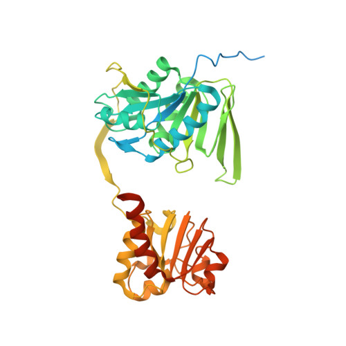
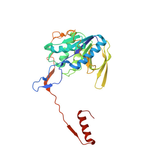
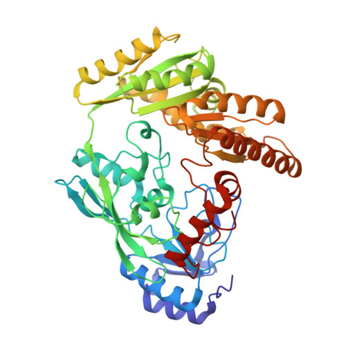
Structure-based electron-confurcation mechanism of the Ldh-EtfAB complex.

Kayastha, K.Katsyv, A.Himmrich, C.Welsch, S.Schuller, J.M.Ermler, U.Muller, V.

(2022) Elife 11

  • DOI: https://doi.org/10.7554/eLife.77095
  • Primary Citation of Related Structures:  
    7QH2

  • PubMed Abstract: 

    Lactate oxidation with NAD + as electron acceptor is a highly endergonic reaction. Some anaerobic bacteria overcome the energetic hurdle by flavin-based electron bifurcation/confurcation (FBEB/FBEC) using a lactate dehydrogenase (Ldh) in concert with the electron-transferring proteins EtfA and EtfB. The electron cryo-microscopically characterized (Ldh-EtfAB) 2 complex of Acetobacterium woodii at 2.43 Å resolution consists of a mobile EtfAB shuttle domain located between the rigid central Ldh and the peripheral EtfAB base units. The FADs of Ldh and the EtfAB shuttle domain contact each other thereby forming the D (dehydrogenation-connected) state. The intermediary Glu37 and Glu139 may harmonize the redox potentials between the FADs and the pyruvate/lactate pair crucial for FBEC. By integrating Alphafold2 calculations a plausible novel B (bifurcation-connected) state was obtained allowing electron transfer between the EtfAB base and shuttle FADs. Kinetic analysis of enzyme variants suggests a correlation between NAD + binding site and D-to-B-state transition implicating a 75° rotation of the EtfAB shuttle domain. The FBEC inactivity when truncating the ferredoxin domain of EtfA substantiates its role as redox relay. Lactate oxidation in Ldh is assisted by the catalytic base His423 and a metal center. On this basis, a comprehensive catalytic mechanism of the FBEC process was proposed.


  • Organizational Affiliation

    Departments of Molecular Membrane Biology of the Max-Planck-Institut for Biophysics, Frankfurt am Main, Germany.


Macromolecules
Find similar proteins by:  (by identity cutoff)  |  3D Structure
Entity ID: 1
MoleculeChains Sequence LengthOrganismDetailsImage
Lactate dehydrogenase (NAD(+),ferredoxin) subunit LctC
A, D
418Acetobacterium woodiiMutation(s): 0 
Gene Names: lctCetfAAwo_c08720
EC: 1.3.1.110
UniProt
Find proteins for H6LBB1 (Acetobacterium woodii (strain ATCC 29683 / DSM 1030 / JCM 2381 / KCTC 1655 / WB1))
Explore H6LBB1 
Go to UniProtKB:  H6LBB1
Entity Groups  
Sequence Clusters30% Identity50% Identity70% Identity90% Identity95% Identity100% Identity
UniProt GroupH6LBB1
Sequence Annotations
Expand
  • Reference Sequence
Find similar proteins by:  (by identity cutoff)  |  3D Structure
Entity ID: 2
MoleculeChains Sequence LengthOrganismDetailsImage
Lactate dehydrogenase (NAD(+),ferredoxin) subunit LctB
B, E
265Acetobacterium woodiiMutation(s): 0 
Gene Names: lctBetfBAwo_c08710
EC: 1.3.1.110
UniProt
Find proteins for H6LBB0 (Acetobacterium woodii (strain ATCC 29683 / DSM 1030 / JCM 2381 / KCTC 1655 / WB1))
Explore H6LBB0 
Go to UniProtKB:  H6LBB0
Entity Groups  
Sequence Clusters30% Identity50% Identity70% Identity90% Identity95% Identity100% Identity
UniProt GroupH6LBB0
Sequence Annotations
Expand
  • Reference Sequence
Find similar proteins by:  (by identity cutoff)  |  3D Structure
Entity ID: 3
MoleculeChains Sequence LengthOrganismDetailsImage
Lactate dehydrogenase (NAD(+),ferredoxin) subunit LctD
C, F
467Acetobacterium woodiiMutation(s): 0 
Gene Names: lctDglcDAwo_c08730
EC: 1.3.1.110
UniProt
Find proteins for H6LBS1 (Acetobacterium woodii (strain ATCC 29683 / DSM 1030 / JCM 2381 / KCTC 1655 / WB1))
Explore H6LBS1 
Go to UniProtKB:  H6LBS1
Entity Groups  
Sequence Clusters30% Identity50% Identity70% Identity90% Identity95% Identity100% Identity
UniProt GroupH6LBS1
Sequence Annotations
Expand
  • Reference Sequence
Experimental Data & Validation

Experimental Data

  • Method: ELECTRON MICROSCOPY
  • Resolution: 2.43 Å
  • Aggregation State: PARTICLE 
  • Reconstruction Method: SINGLE PARTICLE 
EM Software:
TaskSoftware PackageVersion
MODEL REFINEMENTCoot0.9
RECONSTRUCTIONRELION3.1

Structure Validation

View Full Validation Report


Entry History & Funding Information

Deposition Data


Funding OrganizationLocationGrant Number
Max Planck SocietyGermany--

Revision History  (Full details and data files)

  • Version 1.0: 2022-06-29
    Type: Initial release
  • Version 1.1: 2022-07-06
    Changes: Database references