Plant vernalization proteins contain unusual PHD superdomains without histone H3 binding activity.
Franco-Echevarria, E., Rutherford, T.J., Fiedler, M., Dean, C., Bienz, M.(2022) J Biological Chem 298: 102540-102540
- PubMed: 36174674 Search on PubMedSearch on PubMed Central
- DOI: https://doi.org/10.1016/j.jbc.2022.102540
- Primary Citation Related Structures:
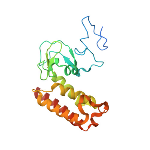
7QCE - PubMed Abstract:
PHD fingers are modular domains in chromatin-associated proteins that decode the methylation status of histone H3 tails. A PHD finger signature is found in plant vernalization (VEL) proteins, which function as accessory factors of the Polycomb system to control flowering in Arabidopsis through an epigenetic silencing mechanism. It has been proposed that VEL PHD fingers bind to methylated histone H3 tails to facilitate association of the Polycomb silencing machinery with target genes. Here, we use structural analysis by X-ray crystallography to show that the VEL PHD finger forms the central module of a larger compact tripartite superdomain that also contains a zinc finger and a four-helix bundle. This PHD superdomain fold is only found in one other family, the OBERON proteins, which have multiple functions in Arabidopsis meristems to control plant growth. The putative histone-binding surface of OBERON proteins exhibits the characteristic three-pronged pocket of histone-binding PHD fingers and binds to methylated histone H3 tails. However, that of VEL PHD fingers lacks this architecture and exhibits unusually high positive surface charge. This VEL PHD superdomain neither binds to unmodified nor variously modified histone H3 tails, as demonstrated by isothermal calorimetry and NMR spectroscopy. Instead, the VEL PHD superdomain interacts with negatively charged polymers. Our evidence argues for evolution of a divergent function for the PHD superdomain in vernalization that does not involve histone tail decoding.
- MRC Laboratory of Molecular Biology, Cambridge, United Kingdom.
Organizational Affiliation: