7QAC | pdb_00007qac

The T2 structure of polycrystalline cubic human insulin


Experimental Data Snapshot

  • Method: POWDER DIFFRACTION
  • Resolution: 2.29 Å

Starting Model: experimental
View more details

wwPDB Validation 3D Report Full Report

Validation slider image for 7QAC

This is version 1.4 of the entry. See complete history

Literature

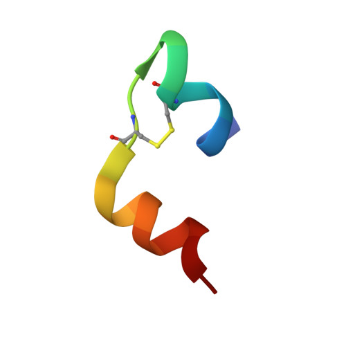
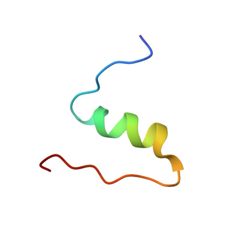
The T 2 structure of polycrystalline cubic human insulin.

Triandafillidis, D.P.Karavassili, F.Spiliopoulou, M.Valmas, A.Athanasiadou, M.Nikolaras, G.Fili, S.Kontou, P.Bowler, M.W.Chasapis, C.T.Von Dreele, R.B.Fitch, A.N.Margiolaki, I.

(2023) Acta Crystallogr D Struct Biol 79: 374-386

  • DOI: https://doi.org/10.1107/S2059798323001328
  • Primary Citation Related Structures: 
    7QAC

  • PubMed Abstract: 

    The polymorphism of human insulin upon pH variation was characterized via X-ray powder diffraction, employing a crystallization protocol previously established for co-crystallization with phenolic derivatives. Two distinct rhombohedral (R3) polymorphs and one cubic (I2 1 3) polymorph were identified with increasing pH, corresponding to the T 6 , T 3 R 3 f and T 2 conformations of insulin, respectively. The structure of the cubic T 2 polymorph was determined via multi-profile stereochemically restrained Rietveld refinement at 2.7 Å resolution. This constitutes the first cubic insulin structure to be determined from crystals grown in the presence of zinc ions, although no zinc binding was observed. The differences of the polycrystalline variant from other cubic insulin structures, as well as the nature of the pH-driven phase transitions, are discussed in detail.


  • Organizational Affiliation
    • Section of Genetics, Cell Biology and Development, Department of Biology, University of Patras, 26504 Patras, Greece.

Macromolecule Content 

  • Total Structure Weight: 5.82 kDa 
  • Atom Count: 467 
  • Modeled Residue Count: 51 
  • Deposited Residue Count: 51 
  • Unique protein chains: 2

Macromolecules

Find similar proteins by:  Sequence   |   3D Structure  
Entity ID: 1
MoleculeChains  Sequence LengthOrganismDetailsImage
Insulin A chain21Homo sapiensMutation(s): 0 
Gene Names: INS
UniProt & NIH Common Fund Data Resources
Find proteins for P01308 (Homo sapiens)
Explore P01308 
Go to UniProtKB:  P01308
PHAROS:  P01308
GTEx:  ENSG00000254647 
Entity Groups
Sequence Clusters30% Identity50% Identity70% Identity90% Identity95% Identity100% Identity
UniProt GroupP01308
Sequence Annotations
Expand
Reference Sequence
Find similar proteins by:|  3D Structure
Entity ID: 2
MoleculeChains  Sequence LengthOrganismDetailsImage
Insulin B chain30Homo sapiensMutation(s): 0 
Gene Names: INS
UniProt & NIH Common Fund Data Resources
Find proteins for P01308 (Homo sapiens)
Explore P01308 
Go to UniProtKB:  P01308
PHAROS:  P01308
GTEx:  ENSG00000254647 
Entity Groups
Sequence Clusters30% Identity50% Identity70% Identity90% Identity95% Identity100% Identity
UniProt GroupP01308
Sequence Annotations
Expand
Reference Sequence

Experimental Data & Validation

Experimental Data

  • Method: POWDER DIFFRACTION
  • Resolution: 2.29 Å
Space Group: I 21 3
Unit Cell:
Length ( Å )Angle ( ˚ )
a = 78.86015α = 90
b = 78.86015β = 90
c = 78.86015γ = 90
Software Package:
Software NamePurpose
GSASrefinement
PRODDdata reduction
MOLREPphasing
Cootmodel building

Structure Validation

View Full Validation Report


Entry History 

& Funding Information

Deposition Data


Funding OrganizationLocationGrant Number
General Secretariat for Research and Technology (GSRT)European Union--
Hellenic Foundation for Research and Innovation (HFRI)Greece3051

Revision History  (Full details and data files)

  • Version 1.0: 2023-06-21
    Type: Initial release
  • Version 1.1: 2023-07-19
    Changes: Data collection
  • Version 1.2: 2023-08-16
    Changes: Data collection
  • Version 1.3: 2024-02-07
    Changes: Refinement description
  • Version 1.4: 2024-10-09
    Changes: Structure summary