7QAA

Crystal structure of RARalpha/RXRalpha ligand binding domain heterodimer in complex with BMS614 and oleic acid


Experimental Data Snapshot

  • Method: X-RAY DIFFRACTION
  • Resolution: 2.76 Å
  • R-Value Free: 0.245 
  • R-Value Work: 0.195 
  • R-Value Observed: 0.197 

wwPDB Validation   3D Report Full Report


Ligand Structure Quality Assessment 


This is version 1.1 of the entry. See complete history


Literature

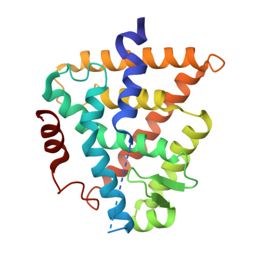
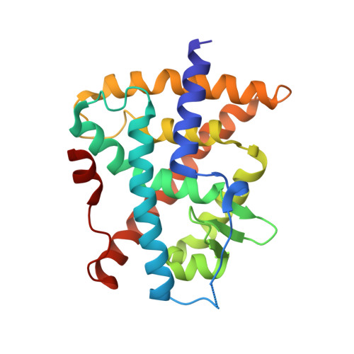
Design and in vitro characterization of RXR variants as tools to investigate the biological role of endogenous rexinoids.

le Maire, A.Rey, M.Vivat, V.Guee, L.Blanc, P.Malosse, C.Chamot-Rooke, J.Germain, P.Bourguet, W.

(2022) J Mol Endocrinol 69: 377-390

  • DOI: https://doi.org/10.1530/JME-22-0021
  • Primary Citation of Related Structures:  
    7PDQ, 7PDT, 7QAA

  • PubMed Abstract: 

    Retinoid X receptors (RXRα, β, and γ) are essential members of the nuclear receptor (NR) superfamily of ligand-dependent transcriptional regulators that bind DNA response elements and control the expression of large gene networks. As obligate heterodimerization partners of many NRs, RXRs are involved in a variety of pathophysiological processes. However, despite this central role in NR signaling, there is still no consensus regarding the precise biological functions of RXRs and the putative role of the endogenous ligands (rexinoids) previously proposed for these receptors. Based on available crystal structures, we introduced a series of amino acid substitutions into the ligand-binding pocket of all three RXR subtypes in order to alter their binding properties. Subsequent characterization using a battery of cell-based and in vitro assays led to the identification of a double mutation abolishing the binding of any ligand while keeping the other receptor functions intact and a triple mutation that selectively impairs interaction with natural rexinoids but not with some synthetic ligands. We also report crystal structures that help understand the specific ligand-binding capabilities of both variants. These RXR variants, either fully disabled for ligand binding or retaining the property of being activated by synthetic compounds, represent unique tools that could be used in future studies to probe the presence of active endogenous rexinoids in tissues/organs and to investigate their role in vivo. Last, we provide data suggesting a possible involvement of fatty acids in the weak interaction of RXRs with corepressors.


  • Organizational Affiliation

    CBS (Centre de Biologie Structurale), Univ Montpellier, CNRS, Inserm, Montpellier, France.


Macromolecules
Find similar proteins by:  (by identity cutoff)  |  3D Structure
Entity ID: 1
MoleculeChains Sequence LengthOrganismDetailsImage
Retinoic acid receptor RXR-alpha233Mus musculusMutation(s): 0 
Gene Names: RxraNr2b1
UniProt
Find proteins for P28700 (Mus musculus)
Explore P28700 
Go to UniProtKB:  P28700
Entity Groups  
Sequence Clusters30% Identity50% Identity70% Identity90% Identity95% Identity100% Identity
UniProt GroupP28700
Sequence Annotations
Expand
  • Reference Sequence
Find similar proteins by:  (by identity cutoff)  |  3D Structure
Entity ID: 2
MoleculeChains Sequence LengthOrganismDetailsImage
Isoform Alpha-1-deltaBC of Retinoic acid receptor alpha235Homo sapiensMutation(s): 0 
Gene Names: RARANR1B1
UniProt & NIH Common Fund Data Resources
Find proteins for P10276 (Homo sapiens)
Explore P10276 
Go to UniProtKB:  P10276
PHAROS:  P10276
GTEx:  ENSG00000131759 
Entity Groups  
Sequence Clusters30% Identity50% Identity70% Identity90% Identity95% Identity100% Identity
UniProt GroupP10276
Sequence Annotations
Expand
  • Reference Sequence
Small Molecules
Ligands 2 Unique
IDChains Name / Formula / InChI Key2D Diagram3D Interactions
BMS (Subject of Investigation/LOI)
Query on BMS

Download Ideal Coordinates CCD File 
D [auth B]4-[(4,4-DIMETHYL-1,2,3,4-TETRAHYDRO-[1,2']BINAPTHALENYL-7-CARBONYL)-AMINO]-BENZOIC ACID
C29 H26 N2 O3
WBEIHCAWTAWTBK-UHFFFAOYSA-N
OLA (Subject of Investigation/LOI)
Query on OLA

Download Ideal Coordinates CCD File 
C [auth A]OLEIC ACID
C18 H34 O2
ZQPPMHVWECSIRJ-KTKRTIGZSA-N
Experimental Data & Validation

Experimental Data

  • Method: X-RAY DIFFRACTION
  • Resolution: 2.76 Å
  • R-Value Free: 0.245 
  • R-Value Work: 0.195 
  • R-Value Observed: 0.197 
  • Space Group: P 61 2 2
Unit Cell:
Length ( Å )Angle ( ˚ )
a = 116.6α = 90
b = 116.6β = 90
c = 207.8γ = 120
Software Package:
Software NamePurpose
PHENIXrefinement
DENZOdata reduction
SCALEPACKdata scaling

Structure Validation

View Full Validation Report


Ligand Structure Quality Assessment 


Entry History & Funding Information

Deposition Data


Funding OrganizationLocationGrant Number
Not funded--

Revision History  (Full details and data files)

  • Version 1.0: 2022-10-05
    Type: Initial release
  • Version 1.1: 2024-01-31
    Changes: Data collection, Refinement description