7Q55

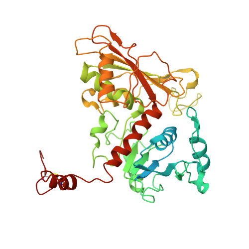
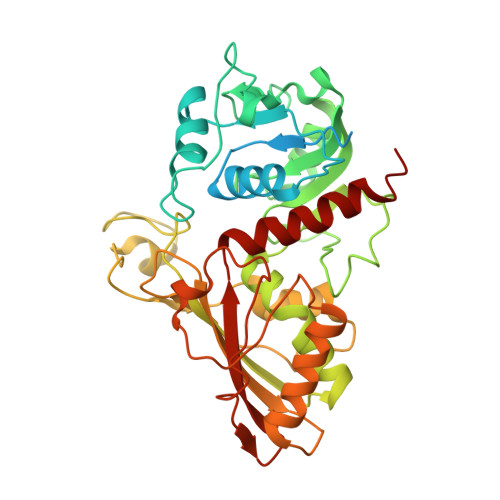
Single Particle Cryo-EM structure of photosynthetic A8B8 glyceraldehyde-3-phosphate dehydrogenase hexadecamer (major conformer) from Spinacia oleracia.


Experimental Data Snapshot

  • Method: ELECTRON MICROSCOPY
  • Resolution: 5.70 Å
  • Aggregation State: PARTICLE 
  • Reconstruction Method: SINGLE PARTICLE 

wwPDB Validation   3D Report Full Report


This is version 1.0 of the entry. See complete history


Literature

Unravelling the regulation pathway of photosynthetic AB-GAPDH.

Marotta, R.Del Giudice, A.Gurrieri, L.Fanti, S.Swuec, P.Galantini, L.Falini, G.Trost, P.Fermani, S.Sparla, F.

(2022) Acta Crystallogr D Struct Biol 78: 1399-1411

  • DOI: https://doi.org/10.1107/S2059798322010014
  • Primary Citation of Related Structures:  
    7Q53, 7Q54, 7Q55, 7Q56, 7Q57

  • PubMed Abstract: 

    Oxygenic phototrophs perform carbon fixation through the Calvin-Benson cycle. Different mechanisms adjust the cycle and the light-harvesting reactions to rapid environmental changes. Photosynthetic glyceraldehyde 3-phosphate dehydrogenase (GAPDH) is a key enzyme in the cycle. In land plants, different photosynthetic GAPDHs exist: the most abundant isoform is formed by A 2 B 2 heterotetramers and the least abundant by A 4 homotetramers. Regardless of the subunit composition, GAPDH is the major consumer of photosynthetic NADPH and its activity is strictly regulated. While A 4 -GAPDH is regulated by CP12, AB-GAPDH is autonomously regulated through the C-terminal extension (CTE) of its B subunits. Reversible inhibition of AB-GAPDH occurs via the oxidation of a cysteine pair located in the CTE and the substitution of NADP(H) with NAD(H) in the cofactor-binding site. These combined conditions lead to a change in the oligomerization state and enzyme inhibition. SEC-SAXS and single-particle cryo-EM analysis were applied to reveal the structural basis of this regulatory mechanism. Both approaches revealed that spinach (A 2 B 2 ) n -GAPDH oligomers with n = 1, 2, 4 and 5 co-exist in a dynamic system. B subunits mediate the contacts between adjacent tetramers in A 4 B 4 and A 8 B 8 oligomers. The CTE of each B subunit penetrates into the active site of a B subunit of the adjacent tetramer, which in turn moves its CTE in the opposite direction, effectively preventing the binding of the substrate 1,3-bisphosphoglycerate in the B subunits. The whole mechanism is made possible, and eventually controlled, by pyridine nucleotides. In fact, NAD(H), by removing NADP(H) from A subunits, allows the entrance of the CTE into the active site of the B subunit, hence stabilizing inhibited oligomers.


  • Organizational Affiliation

    Electron Microscopy Facility (EMF), Italian Institute of Technology (IIT), 16163 Genova, Italy.


Macromolecules
Find similar proteins by:  (by identity cutoff)  |  3D Structure
Entity ID: 1
MoleculeChains Sequence LengthOrganismDetailsImage
Glyceraldehyde-3-phosphate dehydrogenase B, chloroplastic451Spinacia oleraceaMutation(s): 0 
EC: 1.2.1.13
UniProt
Find proteins for P12860 (Spinacia oleracea)
Explore P12860 
Go to UniProtKB:  P12860
Entity Groups  
Sequence Clusters30% Identity50% Identity70% Identity90% Identity95% Identity100% Identity
UniProt GroupP12860
Sequence Annotations
Expand
  • Reference Sequence
Find similar proteins by:  (by identity cutoff)  |  3D Structure
Entity ID: 2
MoleculeChains Sequence LengthOrganismDetailsImage
Glyceraldehyde-3-phosphate dehydrogenase A, chloroplastic402Spinacia oleraceaMutation(s): 0 
EC: 1.2.1.13
UniProt
Find proteins for P19866 (Spinacia oleracea)
Explore P19866 
Go to UniProtKB:  P19866
Entity Groups  
Sequence Clusters30% Identity50% Identity70% Identity90% Identity95% Identity100% Identity
UniProt GroupP19866
Sequence Annotations
Expand
  • Reference Sequence
Small Molecules
Ligands 1 Unique
IDChains Name / Formula / InChI Key2D Diagram3D Interactions
NAD (Subject of Investigation/LOI)
Query on NAD

Download Ideal Coordinates CCD File 
AA [auth G]
BA [auth H]
CA [auth I]
DA [auth J]
EA [auth K]
AA [auth G],
BA [auth H],
CA [auth I],
DA [auth J],
EA [auth K],
FA [auth L],
Q [auth O],
R [auth P],
S [auth Q],
T [auth R],
U [auth A],
V [auth B],
W [auth C],
X [auth D],
Y [auth E],
Z [auth F]
NICOTINAMIDE-ADENINE-DINUCLEOTIDE
C21 H27 N7 O14 P2
BAWFJGJZGIEFAR-NNYOXOHSSA-N
Experimental Data & Validation

Experimental Data

  • Method: ELECTRON MICROSCOPY
  • Resolution: 5.70 Å
  • Aggregation State: PARTICLE 
  • Reconstruction Method: SINGLE PARTICLE 

Structure Validation

View Full Validation Report


Entry History & Funding Information

Deposition Data


Funding OrganizationLocationGrant Number
Grenoble Instruct-ERIC Center (ISBG)FrancePID 1829

Revision History  (Full details and data files)

  • Version 1.0: 2022-11-16
    Type: Initial release