7Q16

Human 14-3-3 zeta fused to the BAD peptide including phosphoserine-74


Experimental Data Snapshot

  • Method: X-RAY DIFFRACTION
  • Resolution: 2.36 Å
  • R-Value Free: 0.241 
  • R-Value Work: 0.197 
  • R-Value Observed: 0.199 

wwPDB Validation   3D Report Full Report


This is version 1.2 of the entry. See complete history


Literature

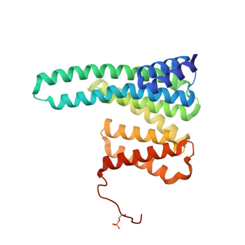
Crystal structure of human 14-3-3 zeta complexed with the noncanonical phosphopeptide from proapoptotic BAD.

Sluchanko, N.N.Tugaeva, K.V.Gushchin, I.Remeeva, A.Kovalev, K.Cooley, R.B.

(2021) Biochem Biophys Res Commun 583: 100-105

  • DOI: https://doi.org/10.1016/j.bbrc.2021.10.053
  • Primary Citation of Related Structures:  
    7Q16

  • PubMed Abstract: 

    Several signaling pathways control phosphorylation of the proapoptotic protein BAD and its phosphorylation-dependent association with 14-3-3 proteins in the cytoplasm. The stability of the 14-3-3/BAD complex determines the cell fate: unphosphorylated BAD escapes from 14-3-3, migrates to the mitochondria and initiates apoptosis. While the 14-3-3/BAD interaction represents a promising drug target, it lacks structural characterization. Among several phosphosites identified in vivo, Ser75 and Ser99 of human BAD match the consensus sequence RXXpSXP recognized by 14-3-3 and, therefore, represent canonical 14-3-3-binding sites. Yet, BAD contains other serines phosphorylatable in vivo, whose role is less understood. Here, we report a 2.36 Å crystal structure of 14-3-3ζ complexed with a BAD fragment which includes residues Ser74 and Ser75, both being substrates for protein kinases. While the BAD peptide is anchored to 14-3-3 by phosphoserine as expected, the BAD peptide was unexpectedly phosphorylated at Ser74 instead of Ser75, revealing noncanonical binding within the amphipathic groove and leading to a one-step positional shift and reorganization of the interface. This observation exemplifies plasticity of the amphipathic 14-3-3 groove in accommodating various peptides and suggests the redundancy of Ser74 and Ser75 phosphosites with respect to binding of BAD to 14-3-3.


  • Organizational Affiliation

    A.N. Bach Institute of Biochemistry, Federal Research Center of Biotechnology of the Russian Academy of Sciences, 119071, Moscow, Russia. Electronic address: nikolai.sluchanko@mail.ru.


Macromolecules
Find similar proteins by:  (by identity cutoff)  |  3D Structure
Entity ID: 1
MoleculeChains Sequence LengthOrganismDetailsImage
14-3-3 protein zeta/delta,Bcl2-associated agonist of cell death244Homo sapiensMutation(s): 4 
Gene Names: YWHAZBADBBC6BCL2L8
UniProt & NIH Common Fund Data Resources
Find proteins for Q92934 (Homo sapiens)
Explore Q92934 
Go to UniProtKB:  Q92934
PHAROS:  Q92934
GTEx:  ENSG00000002330 
Find proteins for P63104 (Homo sapiens)
Explore P63104 
Go to UniProtKB:  P63104
PHAROS:  P63104
GTEx:  ENSG00000164924 
Entity Groups  
Sequence Clusters30% Identity50% Identity70% Identity90% Identity95% Identity100% Identity
UniProt GroupsQ92934P63104
Sequence Annotations
Expand
  • Reference Sequence
Small Molecules
Modified Residues  1 Unique
IDChains TypeFormula2D DiagramParent
SEP
Query on SEP
A
L-PEPTIDE LINKINGC3 H8 N O6 PSER
Experimental Data & Validation

Experimental Data

  • Method: X-RAY DIFFRACTION
  • Resolution: 2.36 Å
  • R-Value Free: 0.241 
  • R-Value Work: 0.197 
  • R-Value Observed: 0.199 
  • Space Group: C 2 2 21
Unit Cell:
Length ( Å )Angle ( ˚ )
a = 85.698α = 90
b = 112.147β = 90
c = 75.719γ = 90
Software Package:
Software NamePurpose
PHENIXrefinement
PDB_EXTRACTdata extraction
XDSdata reduction
STARANISOdata scaling
MOLREPphasing

Structure Validation

View Full Validation Report


Entry History & Funding Information

Deposition Data


Funding OrganizationLocationGrant Number
Russian Science FoundationRussian Federation19-74-10031

Revision History  (Full details and data files)

  • Version 1.0: 2021-11-17
    Type: Initial release
  • Version 1.1: 2022-10-26
    Changes: Derived calculations, Refinement description
  • Version 1.2: 2024-01-31
    Changes: Data collection, Refinement description