7PBD

a1b3 GABA-A receptor + GABA


Experimental Data Snapshot

  • Method: ELECTRON MICROSCOPY
  • Resolution: 3.04 Å
  • Aggregation State: PARTICLE 
  • Reconstruction Method: SINGLE PARTICLE 

wwPDB Validation   3D Report Full Report


This is version 3.0 of the entry. See complete history


Literature

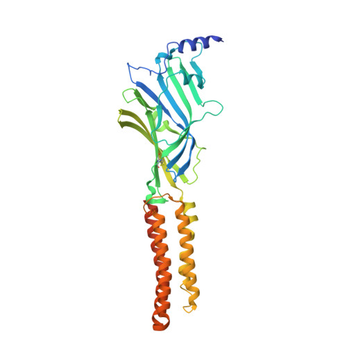
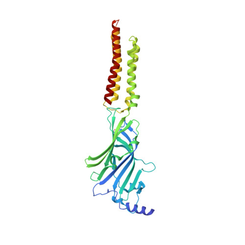
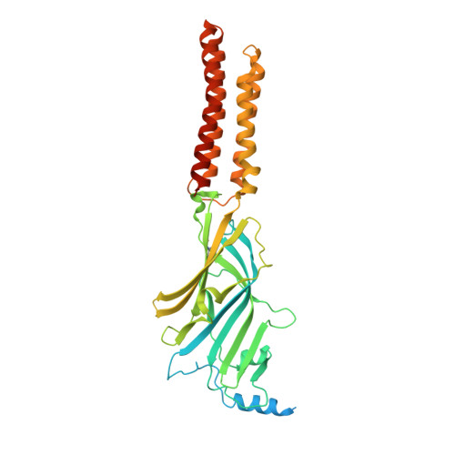
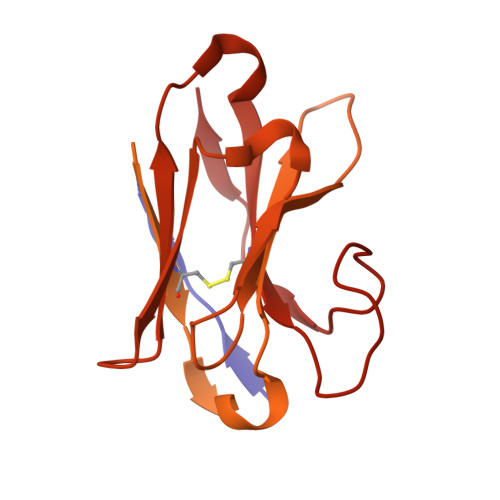
Mechanisms of inhibition and activation of extrasynaptic alpha beta GABA A receptors.

Kasaragod, V.B.Mortensen, M.Hardwick, S.W.Wahid, A.A.Dorovykh, V.Chirgadze, D.Y.Smart, T.G.Miller, P.S.

(2022) Nature 602: 529-533

  • DOI: https://doi.org/10.1038/s41586-022-04402-z
  • Primary Citation of Related Structures:  
    7PBD, 7PBZ, 7PC0

  • PubMed Abstract: 

    Type A GABA (γ-aminobutyric acid) receptors represent a diverse population in the mammalian brain, forming pentamers from combinations of α-, β-, γ-, δ-, ε-, ρ-, θ- and π-subunits 1 . αβ, α4βδ, α6βδ and α5βγ receptors favour extrasynaptic localization, and mediate an essential persistent (tonic) inhibitory conductance in many regions of the mammalian brain 1,2 . Mutations of these receptors in humans are linked to epilepsy and insomnia 3,4 . Altered extrasynaptic receptor function is implicated in insomnia, stroke and Angelman and Fragile X syndromes 1,5 , and drugs targeting these receptors are used to treat postpartum depression 6 . Tonic GABAergic responses are moderated to avoid excessive suppression of neuronal communication, and can exhibit high sensitivity to Zn 2+ blockade, in contrast to synapse-preferring α1βγ, α2βγ and α3βγ receptor responses 5,7-12 . Here, to resolve these distinctive features, we determined structures of the predominantly extrasynaptic αβ GABA A receptor class. An inhibited state bound by both the lethal paralysing agent α-cobratoxin 13 and Zn 2+ was used in comparisons with GABA-Zn 2+ and GABA-bound structures. Zn 2+ nullifies the GABA response by non-competitively plugging the extracellular end of the pore to block chloride conductance. In the absence of Zn 2+ , the GABA signalling response initially follows the canonical route until it reaches the pore. In contrast to synaptic GABA A receptors, expansion of the midway pore activation gate is limited and it remains closed, reflecting the intrinsic low efficacy that characterizes the extrasynaptic receptor. Overall, this study explains distinct traits adopted by αβ receptors that adapt them to a role in tonic signalling.


  • Organizational Affiliation

    Department of Pharmacology, University of Cambridge, Cambridge, UK.


Macromolecules
Find similar proteins by:  (by identity cutoff)  |  3D Structure
Entity ID: 1
MoleculeChains Sequence LengthOrganismDetailsImage
Gamma-aminobutyric acid receptor subunit alpha-1368Homo sapiensMutation(s): 0 
Gene Names: GABRA1
Membrane Entity: Yes 
UniProt & NIH Common Fund Data Resources
Find proteins for P14867 (Homo sapiens)
Explore P14867 
Go to UniProtKB:  P14867
PHAROS:  P14867
GTEx:  ENSG00000022355 
Entity Groups  
Sequence Clusters30% Identity50% Identity70% Identity90% Identity95% Identity100% Identity
UniProt GroupP14867
Sequence Annotations
Expand
  • Reference Sequence
Find similar proteins by:  (by identity cutoff)  |  3D Structure
Entity ID: 2
MoleculeChains Sequence LengthOrganismDetailsImage
Gamma-aminobutyric acid receptor subunit beta-3B,
C,
D [auth E]
451Homo sapiensMutation(s): 0 
Gene Names: GABRB3
Membrane Entity: Yes 
UniProt & NIH Common Fund Data Resources
Find proteins for P28472 (Homo sapiens)
Explore P28472 
Go to UniProtKB:  P28472
PHAROS:  P28472
GTEx:  ENSG00000166206 
Entity Groups  
Sequence Clusters30% Identity50% Identity70% Identity90% Identity95% Identity100% Identity
UniProt GroupP28472
Sequence Annotations
Expand
  • Reference Sequence
Find similar proteins by:  (by identity cutoff)  |  3D Structure
Entity ID: 3
MoleculeChains Sequence LengthOrganismDetailsImage
Gamma-aminobutyric acid receptor subunit alpha-1E [auth D]411Homo sapiensMutation(s): 0 
Gene Names: GABRA1
Membrane Entity: Yes 
UniProt & NIH Common Fund Data Resources
Find proteins for P14867 (Homo sapiens)
Explore P14867 
Go to UniProtKB:  P14867
PHAROS:  P14867
GTEx:  ENSG00000022355 
Entity Groups  
Sequence Clusters30% Identity50% Identity70% Identity90% Identity95% Identity100% Identity
UniProt GroupP14867
Sequence Annotations
Expand
  • Reference Sequence
Find similar proteins by:  (by identity cutoff)  |  3D Structure
Entity ID: 4
MoleculeChains Sequence LengthOrganismDetailsImage
Megabody 25522Lama glamaMutation(s): 0 
Entity Groups  
Sequence Clusters30% Identity50% Identity70% Identity90% Identity95% Identity100% Identity
Sequence Annotations
Expand
  • Reference Sequence
Oligosaccharides

Help

Entity ID: 5
MoleculeChains Length2D Diagram Glycosylation3D Interactions
2-acetamido-2-deoxy-beta-D-glucopyranose-(1-4)-2-acetamido-2-deoxy-beta-D-glucopyranose
G, J
2N-Glycosylation
Glycosylation Resources
GlyTouCan:  G42666HT
GlyCosmos:  G42666HT
GlyGen:  G42666HT
Entity ID: 6
MoleculeChains Length2D Diagram Glycosylation3D Interactions
alpha-D-mannopyranose-(1-3)-[alpha-D-mannopyranose-(1-6)]beta-D-mannopyranose-(1-4)-2-acetamido-2-deoxy-beta-D-glucopyranose-(1-4)-2-acetamido-2-deoxy-beta-D-glucopyranose
H, I, K
5N-Glycosylation
Glycosylation Resources
GlyTouCan:  G22768VO
GlyCosmos:  G22768VO
GlyGen:  G22768VO
Experimental Data & Validation

Experimental Data

  • Method: ELECTRON MICROSCOPY
  • Resolution: 3.04 Å
  • Aggregation State: PARTICLE 
  • Reconstruction Method: SINGLE PARTICLE 

Structure Validation

View Full Validation Report


Entry History & Funding Information

Deposition Data


Funding OrganizationLocationGrant Number
Wellcome TrustUnited Kingdom--

Revision History  (Full details and data files)

  • Version 1.0: 2022-02-09
    Type: Initial release
  • Version 2.0: 2022-02-16
    Changes: Advisory, Atomic model, Data collection, Database references, Derived calculations, Polymer sequence, Source and taxonomy, Structure summary
  • Version 2.1: 2022-02-23
    Changes: Database references
  • Version 2.2: 2022-03-02
    Changes: Database references
  • Version 3.0: 2023-11-15
    Changes: Atomic model, Data collection