7P8P

Crystal structure of Fhit covalently bound to a nucleotide


Experimental Data Snapshot

  • Method: X-RAY DIFFRACTION
  • Resolution: 2.34 Å
  • R-Value Free: 0.246 
  • R-Value Work: 0.193 
  • R-Value Observed: 0.197 

wwPDB Validation   3D Report Full Report


Ligand Structure Quality Assessment 


This is version 1.1 of the entry. See complete history


Literature

Chemical Proteomics of the Tumor Suppressor Fhit Covalently Bound to the Cofactor Ap 3 A Elucidates Its Inhibitory Action on Translation.

Herzog, D.Jansen, J.Missun, M.Diederichs, K.Stengel, F.Marx, A.

(2022) J Am Chem Soc 144: 8613-8623

  • DOI: https://doi.org/10.1021/jacs.2c00815
  • Primary Citation of Related Structures:  
    7P8P

  • PubMed Abstract: 

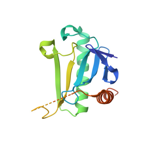
    The tumor suppressor protein fragile histidine triad (Fhit) is known to be associated with genomic instability and apoptosis. The tumor-suppressive function of Fhit depends on the interaction with the alarmone diadenosine triphosphate (Ap 3 A), a noncanonical nucleotide whose concentration increases upon cellular stress. How the Fhit-Ap 3 A complex exerts its signaling function is unknown. Here, guided by a chemical proteomics approach employing a synthetic stable Fhit-Ap 3 A complex, we found that the Fhit-Ap 3 A complex, but not Fhit or Ap 3 A alone, impedes translation. Our findings provide a mechanistic model in which Fhit translocates from the nucleolus into the cytosol upon stress to form an Fhit-Ap 3 A complex. The Fhit-Ap 3 A complex impedes translation both in vitro and in vivo , resulting in reduced cell viability. Overall, our findings provide a mechanistic model by which the tumor suppressor Fhit collaborates with the alarmone Ap 3 A to regulate cellular proliferation.


Macromolecules
Find similar proteins by:  (by identity cutoff)  |  3D Structure
Entity ID: 1
MoleculeChains Sequence LengthOrganismDetailsImage
Bis(5'-adenosyl)-triphosphatase
A, B, C, D
163Homo sapiensMutation(s): 2 
Gene Names: FHIT
EC: 3.6.1.29
UniProt & NIH Common Fund Data Resources
Find proteins for P49789 (Homo sapiens)
Explore P49789 
Go to UniProtKB:  P49789
PHAROS:  P49789
GTEx:  ENSG00000189283 
Entity Groups  
Sequence Clusters30% Identity50% Identity70% Identity90% Identity95% Identity100% Identity
UniProt GroupP49789
Sequence Annotations
Expand
  • Reference Sequence
Small Molecules
Ligands 3 Unique
IDChains Name / Formula / InChI Key2D Diagram3D Interactions
6BI (Subject of Investigation/LOI)
Query on 6BI

Download Ideal Coordinates CCD File 
E [auth A],
G [auth B],
K [auth C],
L [auth D]
[(2~{R},3~{S},4~{R},5~{R})-5-[6-(ethylamino)purin-9-yl]-3,4-bis(oxidanyl)oxolan-2-yl]methyl dihydrogen phosphate
C18 H28 Cl N6 O8 P
QFONDZUXFQHEPG-XKLVTHTNSA-N
TRS
Query on TRS

Download Ideal Coordinates CCD File 
H [auth B]2-AMINO-2-HYDROXYMETHYL-PROPANE-1,3-DIOL
C4 H12 N O3
LENZDBCJOHFCAS-UHFFFAOYSA-O
NA
Query on NA

Download Ideal Coordinates CCD File 
F [auth A],
I [auth B],
J [auth B]
SODIUM ION
Na
FKNQFGJONOIPTF-UHFFFAOYSA-N
Experimental Data & Validation

Experimental Data

  • Method: X-RAY DIFFRACTION
  • Resolution: 2.34 Å
  • R-Value Free: 0.246 
  • R-Value Work: 0.193 
  • R-Value Observed: 0.197 
  • Space Group: P 21 21 21
Unit Cell:
Length ( Å )Angle ( ˚ )
a = 52.422α = 90
b = 61.736β = 90
c = 198.009γ = 90
Software Package:
Software NamePurpose
PHENIXrefinement
PHENIXrefinement
XDSdata reduction
XDSdata scaling
PHENIXphasing

Structure Validation

View Full Validation Report


Ligand Structure Quality Assessment 


Entry History & Funding Information

Deposition Data


Funding OrganizationLocationGrant Number
German Research Foundation (DFG)GermanyMA2288/19

Revision History  (Full details and data files)

  • Version 1.0: 2022-06-01
    Type: Initial release
  • Version 1.1: 2024-01-31
    Changes: Data collection, Refinement description