The Uncommon Active Site of D-Amino Acid Transaminase from Haliscomenobacter hydrossis : Biochemical and Structural Insights into the New Enzyme.
Bakunova, A.K., Nikolaeva, A.Y., Rakitina, T.V., Isaikina, T.Y., Khrenova, M.G., Boyko, K.M., Popov, V.O., Bezsudnova, E.Y.(2021) Molecules 26
- PubMed: 34443642 Search on PubMedSearch on PubMed Central
- DOI: https://doi.org/10.3390/molecules26165053
- Primary Citation Related Structures:
7P7X - PubMed Abstract:
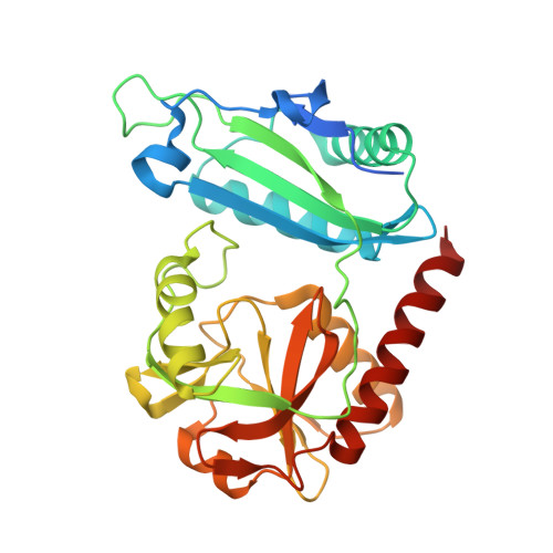
Among industrially important pyridoxal-5'-phosphate (PLP)-dependent transaminases of fold type IV D-amino acid transaminases are the least studied. However, the development of cascade enzymatic processes, including the synthesis of D-amino acids, renewed interest in their study. Here, we describe the identification, biochemical and structural characterization of a new D-amino acid transaminase from Haliscomenobacter hydrossis (Halhy). The new enzyme is strictly specific towards D-amino acids and their keto analogs; it demonstrates one of the highest rates of transamination between D-glutamate and pyruvate. We obtained the crystal structure of the Halhy in the holo form with the protonated Schiff base formed by the K143 and the PLP. Structural analysis revealed a novel set of the active site residues that differ from the key residues forming the active sites of the previously studied D-amino acids transaminases. The active site of Halhy includes three arginine residues, one of which is unique among studied transaminases. We identified critical residues for the Halhy catalytic activity and suggested functions of the arginine residues based on the comparative structural analysis, mutagenesis, and molecular modeling simulations. We suggested a strong positive charge in the O-pocket and the unshaped P-pocket as a structural code for the D-amino acid specificity among transaminases of PLP fold type IV. Characteristics of Halhy complement our knowledge of the structural basis of substrate specificity of D-amino acid transaminases and the sequence-structure-function relationships in these enzymes.
- Bach Institute of Biochemistry, Research Center of Biotechnology of the Russian Academy of Sciences, Leninsky Ave. 33, bld. 2, 119071 Moscow, Russia.
Organizational Affiliation: