Ultra-high resolution X-ray structure of orthorhombic bovine pancreatic Ribonuclease A at 100K.
Lisgarten, D.R., Palmer, R.A., Cooper, J.B., Naylor, C.E., Talbert, R.C., Howlin, B.J., Lisgarten, J.N., Konc, J., Najmudin, S., Lobley, C.M.C.(2023) BMC Chem 17: 91-91
- PubMed: 37501200 Search on PubMedSearch on PubMed Central
- DOI: https://doi.org/10.1186/s13065-023-00959-6
- Primary Citation Related Structures:
7P4R - PubMed Abstract:
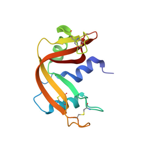
The crystal structure of orthorhombic Bovine Pancreatic Ribonuclease A has been determined to 0.85 Å resolution using low temperature, 100 K, synchrotron X-ray data collected at 16000 keV (λ = 0.77 Å). This is the first ultra-high-resolution structure of a native form of Ribonuclease A to be reported. Refinement carried out with anisotropic displacement parameters, stereochemical restraints, inclusion of H atoms in calculated positions, five [Formula: see text] moieties, eleven ethanol molecules and 293 water molecules, converged with final R values of R1(Free) = 0.129 (4279 reflections) and R1 = 0.112 (85,346 reflections). The refined structure was deposited in the Protein Data Bank as structure 7p4r. Conserved waters, using four high resolution structures, have been investigated. Cluster analysis identified clusters of water molecules that are associated with the active site of Bovine Ribonuclease A. Particular attention has been paid to making detailed comparisons between the present structure and other high quality Bovine Pancreatic Ribonuclease A X-ray crystal structures with special reference to the deposited classic monoclinic structure 3RN3 Howlin et al. (Acta Crystallogr A 45:851-861, 1989). Detailed studies of various aspects of hydrogen bonding and conformation have been carried out with particular reference to active site residues Lys-1, Lys-7, Gln-11, His-12, Lys-41, Asn-44, Thr-45, Lys-66, His-119 and Ser-123. For the two histidine residues in the active site the initial electron density map gives a clear confirmation that the position of His-12 is very similar in the orthorhombic structure to that in 3RN3. In 3RN3 His-119 exhibited poor electron density which was modelled and refined as two distinct sites, A (65%) and B (35%) but with respect to His-119 in the present ultra-high resolution orthorhombic structure there is clear electron density which was modelled and refined as a single conformation distinct from either conformation A or B in 3RN3. Other points of interest include Serine-32 which is disordered at the end of the sidechain in the present orthorhombic form but has been modelled as a single form in 3RN3. Lysine-66: there is density indicating a possible conformation for this residue. However, the density is relatively weak, and the conformation is unclear. Three types of amino acid representation in the ultra-high resolution electron density are examined: (i) sharp with very clearly resolved features, for example Lys-37; (ii) well resolved but clearly divided into two conformations which are well behaved in the refinement, both having high quality geometry, for example Tyr-76; (iii) poor density and difficult or impossible to model, an example is Lys-31 for which density is missing except for Cβ. The side chains of Gln-11, His-12, Lys-41, Thr-45 and His-119 are generally recognised as being closely involved in the enzyme activity. It has also been suggested that Lys-7, Asp-44, Lys-66, Phe-120, Asp-121 and Ser-123 may also have possible roles in this mechanism. A molecular dynamics study on both structures has investigated the conformations of His-119 which was modelled as two conformations in 3RN3 but is observed to have a single clearly defined conformation in the present orthorhombic structure. MD has also been used to investigate Lys-31, Lys-41 and Ser32. The form of the Ribonuclease A enzyme used in both the present study and in 3RN3 (Howlin et al. in Acta Crystallogr A 45:851-861, 1989) includes a sulphate anion which occupies approximately the same location as the [Formula: see text] phosphate group in protein nucleotide complexes (Borkakoti et al. in J Mol Biol 169:743-755, 1983). The present structure contains 5 [Formula: see text] groups SO41151-SO41155 two of which, SO41152 and SO41153 are disordered, SO41152 being in the active site, and 11 EtOH molecules, EOH A 201-EOH A 211 all of which have good geometry. H atoms were built into the EtOH molecules geometrically. Illustrations of these features in the present structure are included here. The sulphates are presumably present in the material purchased for use in the present study. 293 water molecules are included in the present structure compared to 134 in 3RN3 (Howlin et al. in Acta Crystallogr A 45:851-861, 1989).
- Biomolecular Research Group, School of Psychology and Life Sciences, Canterbury Christ Church University, North Holmes Road, Canterbury Kent, CT1 1QU, UK.
Organizational Affiliation: