7P41

Crystal Structure of human mARC1 A165T Variant


Experimental Data Snapshot

  • Method: X-RAY DIFFRACTION
  • Resolution: 1.60 Å
  • R-Value Free: 0.186 
  • R-Value Work: 0.160 
  • R-Value Observed: 0.161 

wwPDB Validation   3D Report Full Report


Ligand Structure Quality Assessment 


This is version 1.3 of the entry. See complete history


Literature

Letter to the editor: The clinically relevant MTARC1 p.Ala165Thr variant impacts neither the fold nor active site architecture of the human mARC1 protein.

Struwe, M.A.Clement, B.Scheidig, A.

(2022) Hepatol Commun 6: 3277-3278

  • DOI: https://doi.org/10.1002/hep4.1984
  • Primary Citation of Related Structures:  
    7P41

  • PubMed Abstract: 

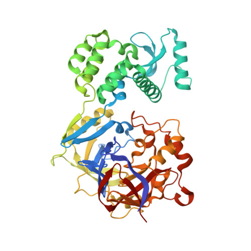
    A study recently published in Hepatology Communications provided insights into a variant of MTARC1 protein, which conveys protection against liver disease. Here, we report a crystal structure of the variant protein at near-atomic resolution and compare it to the structure of the wildtype protein.


  • Organizational Affiliation

    Zoologisches Institut/Strukturbiologie, Christian-Albrechts-Universität zu Kiel, Kiel, Germany.


Macromolecules
Find similar proteins by:  (by identity cutoff)  |  3D Structure
Entity ID: 1
MoleculeChains Sequence LengthOrganismDetailsImage
Mitochondrial amidoxime-reducing component 1,Endolysin,Mitochondrial amidoxime-reducing component 1A [auth D]448Homo sapiensTequatrovirus T4Mutation(s): 0 
Gene Names: MTARC1MARC1MOSC1eT4Tp126
EC: 1.7 (PDB Primary Data), 3.2.1.17 (PDB Primary Data)
UniProt & NIH Common Fund Data Resources
Find proteins for Q5VT66 (Homo sapiens)
Explore Q5VT66 
Go to UniProtKB:  Q5VT66
PHAROS:  Q5VT66
GTEx:  ENSG00000186205 
Find proteins for D9IEF7 (Enterobacteria phage T4)
Explore D9IEF7 
Go to UniProtKB:  D9IEF7
Entity Groups  
Sequence Clusters30% Identity50% Identity70% Identity90% Identity95% Identity100% Identity
UniProt GroupsQ5VT66D9IEF7
Sequence Annotations
Expand
  • Reference Sequence
Small Molecules
Ligands 4 Unique
IDChains Name / Formula / InChI Key2D Diagram3D Interactions
MTE (Subject of Investigation/LOI)
Query on MTE

Download Ideal Coordinates CCD File 
C [auth D]PHOSPHONIC ACIDMONO-(2-AMINO-5,6-DIMERCAPTO-4-OXO-3,7,8A,9,10,10A-HEXAHYDRO-4H-8-OXA-1,3,9,10-TETRAAZA-ANTHRACEN-7-YLMETHYL)ESTER
C10 H14 N5 O6 P S2
HPEUEJRPDGMIMY-IFQPEPLCSA-N
B3P
Query on B3P

Download Ideal Coordinates CCD File 
D
2-[3-(2-HYDROXY-1,1-DIHYDROXYMETHYL-ETHYLAMINO)-PROPYLAMINO]-2-HYDROXYMETHYL-PROPANE-1,3-DIOL
C11 H26 N2 O6
HHKZCCWKTZRCCL-UHFFFAOYSA-N
EFK (Subject of Investigation/LOI)
Query on EFK

Download Ideal Coordinates CCD File 
B [auth D]oxidanyl(oxidanylidene)molybdenum
H Mo O2
VEWPYRVWJVBLDN-UHFFFAOYSA-M
CL
Query on CL

Download Ideal Coordinates CCD File 
E [auth D]CHLORIDE ION
Cl
VEXZGXHMUGYJMC-UHFFFAOYSA-M
Experimental Data & Validation

Experimental Data

  • Method: X-RAY DIFFRACTION
  • Resolution: 1.60 Å
  • R-Value Free: 0.186 
  • R-Value Work: 0.160 
  • R-Value Observed: 0.161 
  • Space Group: P 21 21 21
Unit Cell:
Length ( Å )Angle ( ˚ )
a = 61.063α = 90
b = 74.887β = 90
c = 111.164γ = 90
Software Package:
Software NamePurpose
PHENIXrefinement
XDSdata reduction
Aimlessdata scaling
PHASERphasing
RESOLVEmodel building
PDB_EXTRACTdata extraction

Structure Validation

View Full Validation Report


Ligand Structure Quality Assessment 


Entry History 

Deposition Data

Revision History  (Full details and data files)

  • Version 1.0: 2021-08-18
    Type: Initial release
  • Version 1.1: 2022-05-25
    Changes: Database references
  • Version 1.2: 2022-11-02
    Changes: Database references
  • Version 1.3: 2024-01-31
    Changes: Data collection, Refinement description