7OYO | pdb_00007oyo

Carbonic anhydrase II in complex with Hit4 (MH70)


Experimental Data Snapshot

  • Method: X-RAY DIFFRACTION
  • Resolution: 1.03 Å
  • R-Value Free: 
    0.167 (Depositor), 0.167 (DCC) 
  • R-Value Work: 
    0.136 (Depositor), 0.142 (DCC) 
  • R-Value Observed: 
    0.137 (Depositor) 

Starting Model: experimental
View more details

wwPDB Validation 3D Report Full Report

Validation slider image for 7OYO

Ligand Structure Quality Assessment 


This is version 1.2 of the entry. See complete history

Literature

Identification of specific carbonic anhydrase inhibitors via in situ click chemistry, phage-display and synthetic peptide libraries: comparison of the methods and structural study.

Kugler, M.Hadzima, M.Dzijak, R.Rampmaier, R.Srb, P.Vrzal, L.Voburka, Z.Majer, P.Rezacova, P.Vrabel, M.

(2023) RSC Med Chem 14: 144-153

  • DOI: https://doi.org/10.1039/d2md00330a
  • Primary Citation Related Structures: 
    7OYM, 7OYN, 7OYO, 7OYP, 7OYQ, 7OYR

  • PubMed Abstract: 

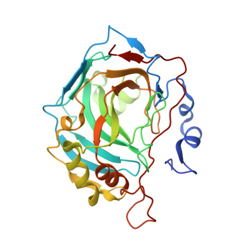
    The development of highly active and selective enzyme inhibitors is one of the priorities of medicinal chemistry. Typically, various high-throughput screening methods are used to find lead compounds from a large pool of synthetic compounds, and these are further elaborated and structurally refined to achieve the desired properties. In an effort to streamline this complex and laborious process, new selection strategies based on different principles have recently emerged as an alternative. Herein, we compare three such selection strategies with the aim of identifying potent and selective inhibitors of human carbonic anhydrase II. All three approaches, in situ click chemistry, phage-display libraries and synthetic peptide libraries, led to the identification of more potent inhibitors when compared to the parent compounds. In addition, one of the inhibitor-peptide conjugates identified from the phage libraries showed greater than 100-fold selectivity for the enzyme isoform used for the compound selection. In an effort to rationalize the binding properties of the conjugates, we performed detailed crystallographic and NMR structural analysis, which revealed the structural basis of the compound affinity towards the enzyme and led to the identification of a novel exosite that could be utilized in the development of isoform specific inhibitors.


  • Organizational Affiliation
    • Institute of Organic Chemistry and Biochemistry of the Czech Academy of Sciences Flemingovo nám. 2 16000 Prague Czech Republic rezacova@uochb.cas.cz vrabel@uochb.cas.cz.

Macromolecule Content 

  • Total Structure Weight: 29.82 kDa 
  • Atom Count: 2,535 
  • Modeled Residue Count: 257 
  • Deposited Residue Count: 260 
  • Unique protein chains: 1

Macromolecules

Find similar proteins by:|  3D Structure
Entity ID: 1
MoleculeChains  Sequence LengthOrganismDetailsImage
Carbonic anhydrase 2260Homo sapiensMutation(s): 0 
Gene Names: CA2
EC: 4.2.1.1 (PDB Primary Data), 4.2.1.69 (UniProt)
UniProt & NIH Common Fund Data Resources
Find proteins for P00918 (Homo sapiens)
Explore P00918 
Go to UniProtKB:  P00918
PHAROS:  P00918
GTEx:  ENSG00000104267 
Entity Groups
Sequence Clusters30% Identity50% Identity70% Identity90% Identity95% Identity100% Identity
UniProt GroupP00918
Sequence Annotations
Expand
Reference Sequence

Small Molecules

Ligands 3 Unique
IDChains Name / Formula / InChI Key2D Diagram3D Interactions
TKR
(Subject of Investigation/LOI)

Query on TKR


Download:Ideal Coordinates CCD File
C [auth A][1-[2-oxidanylidene-2-[2-(4-sulfamoylphenyl)ethylamino]ethyl]-1,2,3-triazolidin-4-yl]methyl hydrogen carbonate
C14 H21 N5 O6 S
UIIITLOQDCCDMB-UHFFFAOYSA-N
DMS

Query on DMS


Download:Ideal Coordinates CCD File
D [auth A]DIMETHYL SULFOXIDE
C2 H6 O S
IAZDPXIOMUYVGZ-UHFFFAOYSA-N
ZN

Query on ZN


Download:Ideal Coordinates CCD File
B [auth A]ZINC ION
Zn
PTFCDOFLOPIGGS-UHFFFAOYSA-N

Experimental Data & Validation

Experimental Data

  • Method: X-RAY DIFFRACTION
  • Resolution: 1.03 Å
  • R-Value Free:  0.167 (Depositor), 0.167 (DCC) 
  • R-Value Work:  0.136 (Depositor), 0.142 (DCC) 
  • R-Value Observed: 0.137 (Depositor) 
Space Group: P 1 21 1
Unit Cell:
Length ( Å )Angle ( ˚ )
a = 42.245α = 90
b = 41.384β = 104.49
c = 72.236γ = 90
Software Package:
Software NamePurpose
XDSdata reduction
XSCALEdata scaling
REFMACrefinement
PDB_EXTRACTdata extraction
FFTphasing

Structure Validation

View Full Validation Report


Ligand Structure Quality Assessment 


Entry History 

Deposition Data

Revision History  (Full details and data files)

  • Version 1.0: 2023-01-18
    Type: Initial release
  • Version 1.1: 2023-02-22
    Changes: Database references
  • Version 1.2: 2024-02-07
    Changes: Data collection, Refinement description