7OMF

Structure of a minimal SF3B core in complex with sudemycin D6 (form I)


Experimental Data Snapshot

  • Method: X-RAY DIFFRACTION
  • Resolution: 3.00 Å
  • R-Value Free: 0.266 
  • R-Value Work: 0.232 
  • R-Value Observed: 0.234 

wwPDB Validation   3D Report Full Report


Ligand Structure Quality Assessment 


This is version 2.0 of the entry. See complete history


Literature

Structural basis of intron selection by U2 snRNP in the presence of covalent inhibitors.

Cretu, C.Gee, P.Liu, X.Agrawal, A.Nguyen, T.V.Ghosh, A.K.Cook, A.Jurica, M.Larsen, N.A.Pena, V.

(2021) Nat Commun 12: 4491-4491

  • DOI: https://doi.org/10.1038/s41467-021-24741-1
  • Primary Citation of Related Structures:  
    7B0I, 7B91, 7B92, 7B9C, 7OMF, 7ONB, 7OPI

  • PubMed Abstract: 

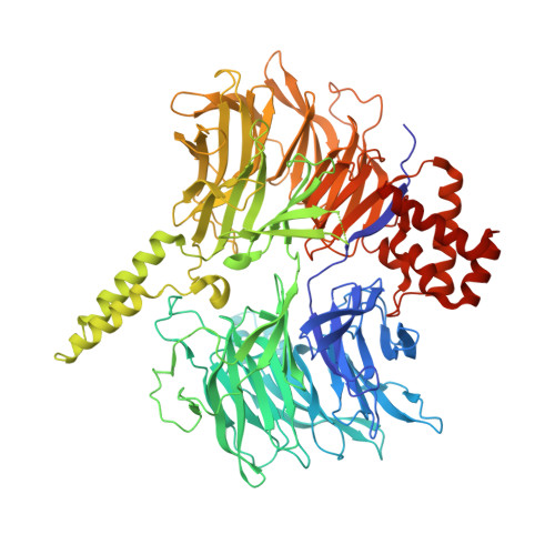
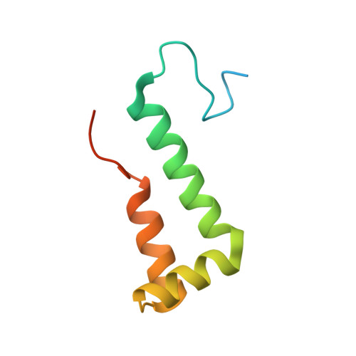
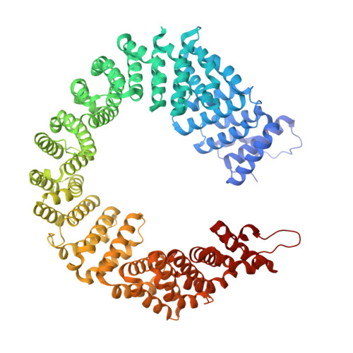
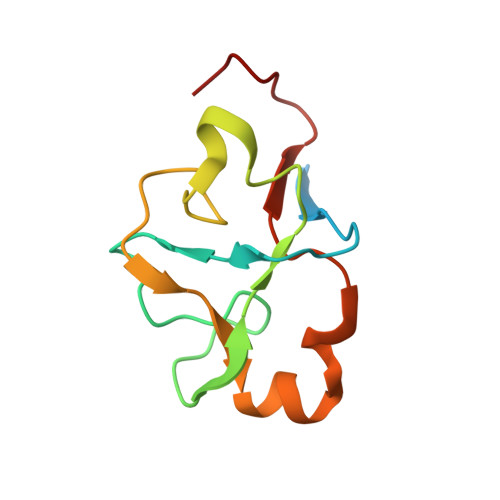
    Intron selection during the formation of prespliceosomes is a critical event in pre-mRNA splicing. Chemical modulation of intron selection has emerged as a route for cancer therapy. Splicing modulators alter the splicing patterns in cells by binding to the U2 snRNP (small nuclear ribonucleoprotein)-a complex chaperoning the selection of branch and 3' splice sites. Here we report crystal structures of the SF3B module of the U2 snRNP in complex with spliceostatin and sudemycin FR901464 analogs, and the cryo-electron microscopy structure of a cross-exon prespliceosome-like complex arrested with spliceostatin A. The structures reveal how modulators inactivate the branch site in a sequence-dependent manner and stall an E-to-A prespliceosome intermediate by covalent coupling to a nucleophilic zinc finger belonging to the SF3B subunit PHF5A. These findings support a mechanism of intron recognition by the U2 snRNP as a toehold-mediated strand invasion and advance an unanticipated drug targeting concept.


  • Organizational Affiliation

    Research Group Mechanisms and Regulation of Splicing, The Institute of Cancer Research, London, UK.


Macromolecules
Find similar proteins by:  (by identity cutoff)  |  3D Structure
Entity ID: 1
MoleculeChains Sequence LengthOrganismDetailsImage
Splicing factor 3B subunit 3899Homo sapiensMutation(s): 0 
Gene Names: SF3B3KIAA0017SAP130
UniProt & NIH Common Fund Data Resources
Find proteins for Q15393 (Homo sapiens)
Explore Q15393 
Go to UniProtKB:  Q15393
PHAROS:  Q15393
GTEx:  ENSG00000189091 
Entity Groups  
Sequence Clusters30% Identity50% Identity70% Identity90% Identity95% Identity100% Identity
UniProt GroupQ15393
Sequence Annotations
Expand
  • Reference Sequence
Find similar proteins by:  (by identity cutoff)  |  3D Structure
Entity ID: 2
MoleculeChains Sequence LengthOrganismDetailsImage
Splicing factor 3B subunit 586Homo sapiensMutation(s): 0 
Gene Names: SF3B5SF3B10
UniProt & NIH Common Fund Data Resources
Find proteins for Q9BWJ5 (Homo sapiens)
Explore Q9BWJ5 
Go to UniProtKB:  Q9BWJ5
PHAROS:  Q9BWJ5
GTEx:  ENSG00000169976 
Entity Groups  
Sequence Clusters30% Identity50% Identity70% Identity90% Identity95% Identity100% Identity
UniProt GroupQ9BWJ5
Sequence Annotations
Expand
  • Reference Sequence
Find similar proteins by:  (by identity cutoff)  |  3D Structure
Entity ID: 3
MoleculeChains Sequence LengthOrganismDetailsImage
Splicing factor 3B subunit 1852Homo sapiensMutation(s): 0 
Gene Names: SF3B1SAP155
UniProt & NIH Common Fund Data Resources
Find proteins for O75533 (Homo sapiens)
Explore O75533 
Go to UniProtKB:  O75533
PHAROS:  O75533
GTEx:  ENSG00000115524 
Entity Groups  
Sequence Clusters30% Identity50% Identity70% Identity90% Identity95% Identity100% Identity
UniProt GroupO75533
Sequence Annotations
Expand
  • Reference Sequence
Find similar proteins by:  (by identity cutoff)  |  3D Structure
Entity ID: 4
MoleculeChains Sequence LengthOrganismDetailsImage
PHD finger-like domain-containing protein 5A108Homo sapiensMutation(s): 0 
Gene Names: PHF5A
UniProt & NIH Common Fund Data Resources
Find proteins for Q7RTV0 (Homo sapiens)
Explore Q7RTV0 
Go to UniProtKB:  Q7RTV0
PHAROS:  Q7RTV0
GTEx:  ENSG00000100410 
Entity Groups  
Sequence Clusters30% Identity50% Identity70% Identity90% Identity95% Identity100% Identity
UniProt GroupQ7RTV0
Sequence Annotations
Expand
  • Reference Sequence
Small Molecules
Experimental Data & Validation

Experimental Data

  • Method: X-RAY DIFFRACTION
  • Resolution: 3.00 Å
  • R-Value Free: 0.266 
  • R-Value Work: 0.232 
  • R-Value Observed: 0.234 
  • Space Group: P 21 21 21
Unit Cell:
Length ( Å )Angle ( ˚ )
a = 106.355α = 90
b = 109.846β = 90
c = 253.3γ = 90
Software Package:
Software NamePurpose
XDSdata reduction
Aimlessdata scaling
PHASERphasing
PHENIXrefinement

Structure Validation

View Full Validation Report


Ligand Structure Quality Assessment 


Entry History & Funding Information

Deposition Data


Funding OrganizationLocationGrant Number
German Research Foundation (DFG)GermanyPE 2079/4-1

Revision History  (Full details and data files)

  • Version 1.0: 2021-08-04
    Type: Initial release
  • Version 2.0: 2024-01-31
    Changes: Data collection, Database references, Non-polymer description, Refinement description