Rational Control of Off-State Heterogeneity in a Photoswitchable Fluorescent Protein Provides Switching Contrast Enhancement.
Adam, V., Hadjidemetriou, K., Jensen, N., Shoeman, R.L., Woodhouse, J., Aquila, A., Banneville, A.S., Barends, T.R.M., Bezchastnov, V., Boutet, S., Byrdin, M., Cammarata, M., Carbajo, S., Eleni Christou, N., Coquelle, N., De la Mora, E., El Khatib, M., Moreno Chicano, T., Bruce Doak, R., Fieschi, F., Foucar, L., Glushonkov, O., Gorel, A., Grunbein, M.L., Hilpert, M., Hunter, M., Kloos, M., Koglin, J.E., Lane, T.J., Liang, M., Mantovanelli, A., Nass, K., Nass Kovacs, G., Owada, S., Roome, C.M., Schiro, G., Seaberg, M., Stricker, M., Thepaut, M., Tono, K., Ueda, K., Uriarte, L.M., You, D., Zala, N., Domratcheva, T., Jakobs, S., Sliwa, M., Schlichting, I., Colletier, J.P., Bourgeois, D., Weik, M.(2022) Chemphyschem 23: e202200192-e202200192
- PubMed: 35959919 Search on PubMedSearch on PubMed Central
- DOI: https://doi.org/10.1002/cphc.202200192
- Primary Citation Related Structures:
7AMB, 7AMF, 7O7C, 7O7D, 7O7E, 7O7H, 7O7U, 7O7V, 7O7W, 7O7X - PubMed Abstract:
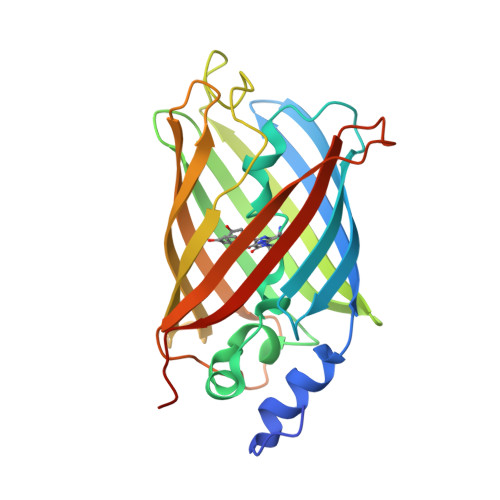
Reversibly photoswitchable fluorescent proteins are essential markers for advanced biological imaging, and optimization of their photophysical properties underlies improved performance and novel applications. Here we establish a link between photoswitching contrast, one of the key parameters that dictate the achievable resolution in nanoscopy applications, and chromophore conformation in the non-fluorescent state of rsEGFP2, a widely employed label in REversible Saturable OpticaL Fluorescence Transitions (RESOLFT) microscopy. Upon illumination, the cis chromophore of rsEGFP2 isomerizes to two distinct off-state conformations, trans1 and trans2, located on either side of the V151 side chain. Reducing or enlarging the side chain at this position (V151A and V151L variants) leads to single off-state conformations that exhibit higher and lower switching contrast, respectively, compared to the rsEGFP2 parent. The combination of structural information obtained by serial femtosecond crystallography with high-level quantum chemical calculations and with spectroscopic and photophysical data determined in vitro suggests that the changes in switching contrast arise from blue- and red-shifts of the absorption bands associated to trans1 and trans2, respectively. Thus, due to elimination of trans2, the V151A variants of rsEGFP2 and its superfolding variant rsFolder2 display a more than two-fold higher switching contrast than their respective parent proteins, both in vitro and in E. coli cells. The application of the rsFolder2-V151A variant is demonstrated in RESOLFT nanoscopy. Our study rationalizes the connection between structural and photophysical chromophore properties and suggests a means to rationally improve fluorescent proteins for nanoscopy applications.
- Univ. Grenoble Alpes, CEA, CNRS, Institut de Biologie Structurale, F-38044, Grenoble, France.
Organizational Affiliation: