


Structure of thaumatin determined at SwissFEL using native-SAD at 5.99 keV with photon energy bandwidth of 0.26%
Nass, K.To be published.
Experimental Data Snapshot
wwPDB Validation 3D Report Full Report

Entity ID: 1 | |||||
|---|---|---|---|---|---|
| Molecule | Chains | Sequence Length | Organism | Details | Image |
| Thaumatin-1 | 207 | Thaumatococcus daniellii | Mutation(s): 0 | 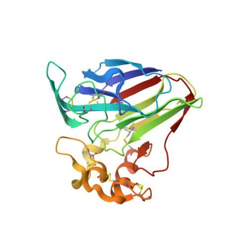 | |
UniProt | |||||
Entity Groups | |||||
| Sequence Clusters | 30% Identity50% Identity70% Identity90% Identity95% Identity100% Identity | ||||
| UniProt Group | P02883 | ||||
Sequence AnnotationsExpand | |||||
Reference Sequence | |||||
| Ligands 1 Unique | |||||
|---|---|---|---|---|---|
| ID | Chains | Name / Formula / InChI Key | 2D Diagram | 3D Interactions | |
| TLA Download:Ideal Coordinates CCD File | B [auth A] | L(+)-TARTARIC ACID C4 H6 O6 FEWJPZIEWOKRBE-JCYAYHJZSA-N |  | ||
| Length ( Å ) | Angle ( ˚ ) |
|---|---|
| a = 58.52 | α = 90 |
| b = 58.52 | β = 90 |
| c = 151.3 | γ = 90 |
| Software Name | Purpose |
|---|---|
| CrystFEL | data collection |
| CrystFEL | data reduction |
| CrystFEL | data scaling |
| SHARP | phasing |
| PHENIX | refinement |
| ARP/wARP | model building |
| BUCCANEER | model building |


