7NXL

Structure of human cathepsin K in complex with the acrylamide inhibitor Gu3110


Experimental Data Snapshot

  • Method: X-RAY DIFFRACTION
  • Resolution: 1.80 Å
  • R-Value Free: 0.209 
  • R-Value Work: 0.175 
  • R-Value Observed: 0.176 

wwPDB Validation   3D Report Full Report


Ligand Structure Quality Assessment 


This is version 2.0 of the entry. See complete history


Literature

An Activity-Based Probe for Cathepsin K Imaging with Excellent Potency and Selectivity.

Lemke, C.Benysek, J.Brajtenbach, D.Breuer, C.Jilkova, A.Horn, M.Busa, M.Ulrychova, L.Illies, A.Kubatzky, K.F.Bartz, U.Mares, M.Gutschow, M.

(2021) J Med Chem 64: 13793-13806

  • DOI: https://doi.org/10.1021/acs.jmedchem.1c01178
  • Primary Citation of Related Structures:  
    7NXL, 7NXM

  • PubMed Abstract: 

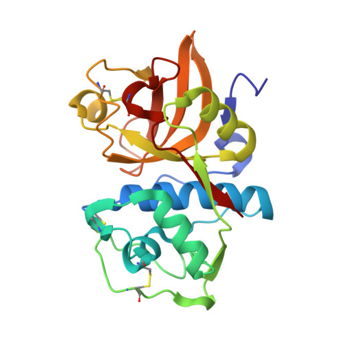
    The cysteine protease cathepsin K is a target for the treatment of diseases associated with high bone turnover. Cathepsin K is mainly expressed in osteoclasts and responsible for the destruction of the proteinaceous components of the bone matrix. We designed various fluorescent activity-based probes (ABPs) and their precursors that bind to and inactivate cathepsin K. ABP 25 exhibited extraordinary potency ( k inac / K i = 35,300 M -1 s -1 ) and selectivity for human cathepsin K. Crystal structures of cathepsin K in complex with ABP 25 and its nonfluorescent precursor 21 were determined to characterize the binding mode of this new type of acrylamide-based Michael acceptor with the particular orientation of the dibenzylamine moiety to the primed subsite region. The cyanine-5 containing probe 25 allowed for sensitive detection of cathepsin K, selective visualization in complex proteomes, and live cell imaging of a human osteosarcoma cell line, underlining its applicability in a pathophysiological environment.


  • Organizational Affiliation

    Pharmaceutical Institute, Pharmaceutical & Medicinal Chemistry, University of Bonn, An der Immenburg 4, Bonn 53121, Germany.


Macromolecules
Find similar proteins by:  (by identity cutoff)  |  3D Structure
Entity ID: 1
MoleculeChains Sequence LengthOrganismDetailsImage
Cathepsin KA [auth AAA]216Homo sapiensMutation(s): 0 
Gene Names: CTSKCTSOCTSO2
EC: 3.4.22.38
UniProt & NIH Common Fund Data Resources
Find proteins for P43235 (Homo sapiens)
Explore P43235 
Go to UniProtKB:  P43235
PHAROS:  P43235
GTEx:  ENSG00000143387 
Entity Groups  
Sequence Clusters30% Identity50% Identity70% Identity90% Identity95% Identity100% Identity
UniProt GroupP43235
Sequence Annotations
Expand
  • Reference Sequence
Small Molecules
Ligands 2 Unique
IDChains Name / Formula / InChI Key2D Diagram3D Interactions
UUK (Subject of Investigation/LOI)
Query on UUK

Download Ideal Coordinates CCD File 
C [auth AAA]tert-butyl (1-((4-(dibenzylamino)-4-oxobutyl)amino)-4-methyl-1-oxopentan-2-yl)carbamate
C29 H41 N3 O4
RIIFVLHFONFLOV-VWLOTQADSA-N
SO4
Query on SO4

Download Ideal Coordinates CCD File 
B [auth AAA]SULFATE ION
O4 S
QAOWNCQODCNURD-UHFFFAOYSA-L
Experimental Data & Validation

Experimental Data

  • Method: X-RAY DIFFRACTION
  • Resolution: 1.80 Å
  • R-Value Free: 0.209 
  • R-Value Work: 0.175 
  • R-Value Observed: 0.176 
  • Space Group: P 2 21 21
Unit Cell:
Length ( Å )Angle ( ˚ )
a = 32.366α = 90
b = 71.819β = 90
c = 80.771γ = 90
Software Package:
Software NamePurpose
REFMACrefinement
XDSdata reduction
XDSdata scaling
MOLREPphasing

Structure Validation

View Full Validation Report


Ligand Structure Quality Assessment 


Entry History 

Deposition Data

Revision History  (Full details and data files)

  • Version 1.0: 2021-09-08
    Type: Initial release
  • Version 2.0: 2021-10-13
    Changes: Atomic model, Data collection, Database references, Non-polymer description