7NMK

Crystal structure of the heterocyclic toxin methyltransferase from Mycobacterium tuberculosis with bound methylation product 1-methoxyquinolin-4(1H)-one


Experimental Data Snapshot

  • Method: X-RAY DIFFRACTION
  • Resolution: 1.20 Å
  • R-Value Free: 0.187 
  • R-Value Work: 0.169 
  • R-Value Observed: 0.170 

wwPDB Validation   3D Report Full Report


This is version 1.3 of the entry. See complete history


Literature

Structural basis of O-methylation of (2-heptyl-)1-hydroxyquinolin-4(1H)-one and related compounds by the heterocyclic toxin methyltransferase Rv0560c of Mycobacterium tuberculosis.

Sartor, P.Denkhaus, L.Gerhardt, S.Einsle, O.Fetzner, S.

(2021) J Struct Biol 213: 107794-107794

  • DOI: https://doi.org/10.1016/j.jsb.2021.107794
  • Primary Citation of Related Structures:  
    7BGG, 7NDM, 7NMK, 7NOY

  • PubMed Abstract: 

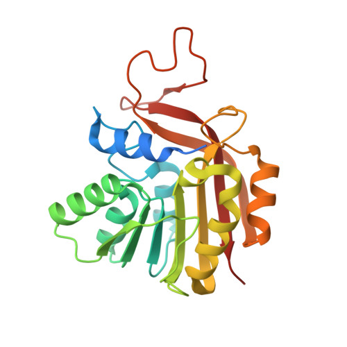
    The S-adenosyl-L-methionine-dependent methyltransferase Rv0560c of Mycobacterium tuberculosis belongs to an orthologous group of heterocyclic toxin methyltransferases (Htm) which likely contribute to resistance of mycobacteria towards antimicrobial natural compounds as well as drugs. Htm M.t. catalyzes the methylation of the Pseudomonas aeruginosa toxin 2-heptyl-1-hydroxyquinolin-4(1H)-one (also known as 2-heptyl-4-hydroxyquinoline N-oxide), a potent inhibitor of respiratory electron transfer, its 1-hydroxyquinolin-4(1H)-one core (QNO), structurally related (iso)quinolones, and some mycobactericidal compounds. In this study, crystal structures of Htm M.t. in complex with S-adenosyl-L-homocysteine (SAH) and the methyl-accepting substrates QNO or 4-hydroxyisoquinoline-1(2H)-one, or the methylated product 1-methoxyquinolin-4(1H)-one, were determined at < 1.9 Å resolution. The monomeric protein exhibits the typical Rossmann fold topology and conserved residues of class I methyltransferases. Its SAH binding pocket is connected via a short tunnel to a large solvent-accessible cavity, which accommodates the methyl-accepting substrate. Residues W44, F168, and F208 in connection with F212 form a hydrophobic clamp around the heteroaromatic ring of the methyl-accepting substrate and likely play a major role in substrate positioning. Structural and biochemical data suggest that H139 and T136 are key active site residues, with H139 acting as general base that activates the methyl-accepting hydroxy group. Our structural data may contribute to the design of Htm inhibitors or of antimycobacterial drugs unamenable for methylation.


  • Organizational Affiliation

    Institute of Molecular Microbiology and Biotechnology, University of Münster, Münster, Germany. Electronic address: sartor@uni-muenster.de.


Macromolecules
Find similar proteins by:  (by identity cutoff)  |  3D Structure
Entity ID: 1
MoleculeChains Sequence LengthOrganismDetailsImage
2-heptyl-1-hydroxyquinolin-4(1H)-one methyltransferase245Mycobacterium tuberculosis H37RvMutation(s): 0 
Gene Names: Rv0560c
UniProt
Find proteins for P9WKL5 (Mycobacterium tuberculosis (strain ATCC 25618 / H37Rv))
Explore P9WKL5 
Go to UniProtKB:  P9WKL5
Entity Groups  
Sequence Clusters30% Identity50% Identity70% Identity90% Identity95% Identity100% Identity
UniProt GroupP9WKL5
Sequence Annotations
Expand
  • Reference Sequence
Small Molecules
Ligands 4 Unique
IDChains Name / Formula / InChI Key2D Diagram3D Interactions
SAH
Query on SAH

Download Ideal Coordinates CCD File 
B [auth A]S-ADENOSYL-L-HOMOCYSTEINE
C14 H20 N6 O5 S
ZJUKTBDSGOFHSH-WFMPWKQPSA-N
UJH (Subject of Investigation/LOI)
Query on UJH

Download Ideal Coordinates CCD File 
C [auth A]1-methoxy-4-oxoquinoline
C10 H9 N O2
QKCKRWUYZIGMET-UHFFFAOYSA-N
FMT
Query on FMT

Download Ideal Coordinates CCD File 
F [auth A],
G [auth A],
H [auth A],
I [auth A]
FORMIC ACID
C H2 O2
BDAGIHXWWSANSR-UHFFFAOYSA-N
NA
Query on NA

Download Ideal Coordinates CCD File 
D [auth A],
E [auth A]
SODIUM ION
Na
FKNQFGJONOIPTF-UHFFFAOYSA-N
Experimental Data & Validation

Experimental Data

  • Method: X-RAY DIFFRACTION
  • Resolution: 1.20 Å
  • R-Value Free: 0.187 
  • R-Value Work: 0.169 
  • R-Value Observed: 0.170 
  • Space Group: P 31 2 1
Unit Cell:
Length ( Å )Angle ( ˚ )
a = 69.866α = 90
b = 69.866β = 90
c = 95.644γ = 120
Software Package:
Software NamePurpose
BUSTERrefinement
XDSdata reduction
Aimlessdata scaling
PHASERphasing

Structure Validation

View Full Validation Report


Entry History & Funding Information

Deposition Data


Funding OrganizationLocationGrant Number
German Research Foundation (DFG)GermanyFE 383/25-1

Revision History  (Full details and data files)

  • Version 1.0: 2021-09-22
    Type: Initial release
  • Version 1.1: 2021-09-29
    Changes: Data collection, Database references
  • Version 1.2: 2021-10-06
    Changes: Data collection, Database references
  • Version 1.3: 2024-01-31
    Changes: Data collection, Refinement description