7N3C | pdb_00007n3c

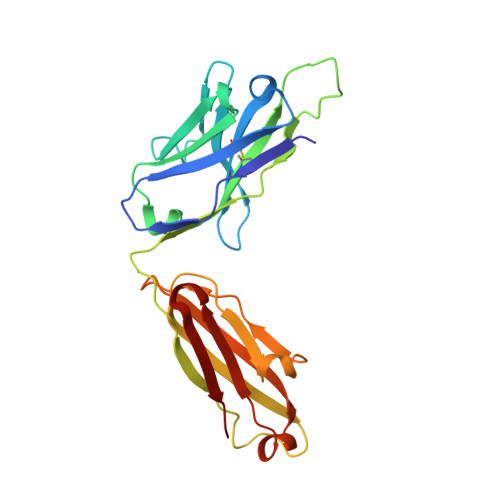
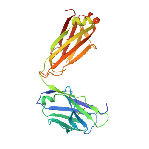
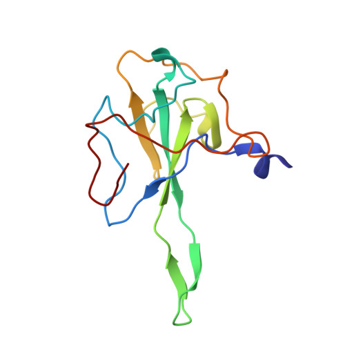
Crystal Structure of Human Fab S24-202 in the complex with the N-terminal Domain of Nucleocapsid protein from SARS CoV-2


Experimental Data Snapshot

  • Method: X-RAY DIFFRACTION
  • Resolution: 1.82 Å
  • R-Value Free: 
    0.210 (Depositor), 0.209 (DCC) 
  • R-Value Work: 
    0.179 (Depositor), 0.179 (DCC) 
  • R-Value Observed: 
    0.181 (Depositor) 

Starting Models: experimental
View more details

wwPDB Validation 3D Report Full Report

Validation slider image for 7N3C

This is version 1.4 of the entry. See complete history. 

Literature

Epitopes recognition of SARS-CoV-2 nucleocapsid RNA binding domain by human monoclonal antibodies.

Kim, Y., Maltseva, N., Tesar, C., Jedrzejczak, R., Endres, M., Ma, H., Dugan, H.L., Stamper, C.T., Chang, C., Li, L., Changrob, S., Zheng, N.Y., Huang, M., Ramanathan, A., Wilson, P., Michalska, K., Joachimiak, A.

(2024) iScience 27: 108976-108976

  • DOI: https://doi.org/10.1016/j.isci.2024.108976
  • Primary Citation Related Structures: 
    6VYO, 6WKP, 7N3C, 7N3D, 7STR, 7STS, 7SUE

  • PubMed Abstract: 

    Coronavirus nucleocapsid protein (NP) of SARS-CoV-2 plays a central role in many functions important for virus proliferation including packaging and protecting genomic RNA. The protein shares sequence, structure, and architecture with nucleocapsid proteins from betacoronaviruses. The N-terminal domain (NP RBD ) binds RNA and the C-terminal domain is responsible for dimerization. After infection, NP is highly expressed and triggers robust host immune response. The anti-NP antibodies are not protective and not neutralizing but can effectively detect viral proliferation soon after infection. Two structures of SARS-CoV-2 NP RBD were determined providing a continuous model from residue 48 to 173, including RNA binding region and key epitopes. Five structures of NP RBD complexes with human mAbs were isolated using an antigen-bait sorting. Complexes revealed a distinct complement-determining regions and unique sets of epitope recognition. This may assist in the early detection of pathogens and designing peptide-based vaccines. Mutations that significantly increase viral load were mapped on developed, full length NP model, likely impacting interactions with host proteins and viral RNA.


  • Organizational Affiliation: 
    • Center for Structural Biology of Infectious Diseases, Consortium for Advanced Science and Engineering, University of Chicago, Chicago, IL 60367, USA.

Macromolecule Content 

  • Total Structure Weight: 64.19 kDa 
  • Atom Count: 4,882 
  • Modeled Residue Count: 570 
  • Deposited Residue Count: 571 
  • Unique protein chains: 3

Macromolecules

Find similar proteins by:|  3D Structure
Entity ID: 1
MoleculeChains  Sequence LengthOrganismDetailsImage
S24-202 Fab heavy chainA [auth H]227Homo sapiensMutation(s): 0 
Entity Groups
Sequence Clusters30% Identity50% Identity70% Identity90% Identity95% Identity100% Identity
Sequence Annotations
Expand
Reference Sequence
Find similar proteins by:|  3D Structure
Entity ID: 2
MoleculeChains  Sequence LengthOrganismDetailsImage
S24-202 Fab light chainB [auth L]214Homo sapiensMutation(s): 0 
Entity Groups
Sequence Clusters30% Identity50% Identity70% Identity90% Identity95% Identity100% Identity
Sequence Annotations
Expand
Reference Sequence
Find similar proteins by:|  3D Structure
Entity ID: 3
MoleculeChains  Sequence LengthOrganismDetailsImage
Nucleoprotein130Severe acute respiratory syndrome coronavirus 2Mutation(s): 0 
UniProt
Find proteins for P0DTC9 (Severe acute respiratory syndrome coronavirus 2)
Explore P0DTC9 
Go to UniProtKB:  P0DTC9
Entity Groups
Sequence Clusters30% Identity50% Identity70% Identity90% Identity95% Identity100% Identity
UniProt GroupP0DTC9
Sequence Annotations
Expand
Reference Sequence

Small Molecules

Ligands 3 Unique
IDChains Name / Formula / InChI Key2D Diagram3D Interactions
IOD

Query on IOD


Download:Ideal Coordinates CCD File
I [auth H]
J [auth H]
K [auth H]
L [auth H]
O [auth L]
I [auth H],
J [auth H],
K [auth H],
L [auth H],
O [auth L],
P [auth L],
Q [auth L],
T [auth C],
U [auth C],
V [auth C],
W [auth C],
X [auth C]
IODIDE ION
I
XMBWDFGMSWQBCA-UHFFFAOYSA-M
PO4

Query on PO4


Download:Ideal Coordinates CCD File
Y [auth C]PHOSPHATE ION
O4 P
NBIIXXVUZAFLBC-UHFFFAOYSA-K
EDO

Query on EDO


Download:Ideal Coordinates CCD File
D [auth H]
E [auth H]
F [auth H]
G [auth H]
H
D [auth H],
E [auth H],
F [auth H],
G [auth H],
H,
M [auth L],
N [auth L],
R [auth C],
S [auth C]
1,2-ETHANEDIOL
C2 H6 O2
LYCAIKOWRPUZTN-UHFFFAOYSA-N

Experimental Data & Validation

Experimental Data

  • Method: X-RAY DIFFRACTION
  • Resolution: 1.82 Å
  • R-Value Free:  0.210 (Depositor), 0.209 (DCC) 
  • R-Value Work:  0.179 (Depositor), 0.179 (DCC) 
  • R-Value Observed: 0.181 (Depositor) 
Space Group: P 1 21 1
Unit Cell:
Length ( Å )Angle ( ˚ )
a = 72.622α = 90
b = 64.02β = 91.091
c = 89.209γ = 90
Software Package:
Software NamePurpose
PHENIXrefinement
HKL-3000data reduction
HKL-3000data scaling
HKL-3000phasing
MOLREPphasing

Structure Validation

View Full Validation Report


Entry History 

& Funding Information

Deposition Data


Funding OrganizationLocationGrant Number
National Institutes of Health/National Institute Of Allergy and Infectious Diseases (NIH/NIAID)United States--

Revision History  (Full details and data files)

  • Version 1.0: 2021-07-07
    Type: Initial release
  • Version 1.1: 2021-07-28
    Changes: Database references, Structure summary
  • Version 1.2: 2023-10-18
    Changes: Data collection, Database references, Refinement description
  • Version 1.3: 2024-02-28
    Changes: Database references
  • Version 1.4: 2024-10-30
    Changes: Structure summary