7MLW | pdb_00007mlw

Burkholderia sp. TJI49 Guanidine-I riboswitch


Experimental Data Snapshot

  • Method: X-RAY DIFFRACTION
  • Resolution: 2.70 Å
  • R-Value Free: 
    0.270 (Depositor), 0.276 (DCC) 
  • R-Value Work: 
    0.218 (Depositor), 0.239 (DCC) 
  • R-Value Observed: 
    0.223 (Depositor) 

Starting Model: experimental
View more details

wwPDB Validation 3D Report Full Report

Validation slider image for 7MLW

Ligand Structure Quality Assessment 


This is version 1.1 of the entry. See complete history

Literature

An uncommon [K + (Mg 2+ ) 2 ] metal ion triad imparts stability and selectivity to the Guanidine-I riboswitch.

Trachman 3rd, R.J.Ferre-D'Amare, A.R.

(2021) RNA 27: 1257-1264

  • DOI: https://doi.org/10.1261/rna.078824.121
  • Primary Citation Related Structures: 
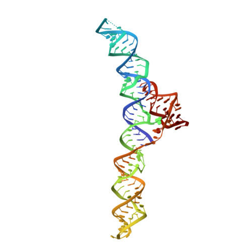
    7MLW

  • PubMed Abstract: 

    The widespread ykkC -I riboswitch class exemplifies divergent riboswitch evolution. To analyze how natural selection has diversified its versatile RNA fold, we determined the X-ray crystal structure of the Burkholderia sp. TJI49 ykkC -I subtype-1 (Guanidine-I) riboswitch aptamer domain. Differing from the previously reported structures of orthologs from Dickeya dadantii and Sulfobacillus acidophilus , our Burkholderia structure reveals a chelated K + ion adjacent to two Mg 2+ ions in the guanidine-binding pocket. Thermal melting analysis shows that K + chelation, which induces localized conformational changes in the binding pocket, improves guanidinium-RNA interactions. Analysis of ribosome structures suggests that the [K + (Mg 2+ ) 2 ] ion triad is uncommon. It is, however, reminiscent of metal ion clusters found in the active sites of ribozymes and DNA polymerases. Previous structural characterization of ykkC -I subtype-2 RNAs, which bind the effector ligands ppGpp and PRPP, indicate that in those paralogs, an adenine responsible for K + chelation in the Burkholderia Guanidine-I riboswitch is replaced by a pyrimidine. This mutation results in a water molecule and Mg 2+ ion binding in place of the K + ion. Thus, our structural analysis demonstrates how ion and solvent chelation tune divergent ligand specificity and affinity among ykkC -I riboswitches.


  • Organizational Affiliation
    • Biochemistry and Biophysics Center, National Heart, Lung, and Blood Institute, Bethesda, Maryland 20892-8012, USA.

Macromolecule Content 

  • Total Structure Weight: 42.27 kDa 
  • Atom Count: 2,748 
  • Modeled Residue Count: 125 
  • Deposited Residue Count: 128 
  • Unique nucleic acid chains: 1

Macromolecules

Find similar nucleic acids by:  (by identity cutoff) 
Entity ID: 1
MoleculeChains LengthOrganismImage
Guanidine-I riboswitchA [auth F]128Burkholderia sp. TJI49
Sequence Annotations
Expand
Reference Sequence

Small Molecules

Ligands 5 Unique
IDChains Name / Formula / InChI Key2D Diagram3D Interactions
PO4

Query on PO4


Download:Ideal Coordinates CCD File
C [auth F],
D [auth F],
E [auth F],
F
PHOSPHATE ION
O4 P
NBIIXXVUZAFLBC-UHFFFAOYSA-K
SR

Query on SR


Download:Ideal Coordinates CCD File
G [auth F],
H [auth F]
STRONTIUM ION
Sr
PWYYWQHXAPXYMF-UHFFFAOYSA-N
GAI
(Subject of Investigation/LOI)

Query on GAI


Download:Ideal Coordinates CCD File
B [auth F]GUANIDINE
C H5 N3
ZRALSGWEFCBTJO-UHFFFAOYSA-N
K
(Subject of Investigation/LOI)

Query on K


Download:Ideal Coordinates CCD File
L [auth F],
M [auth F],
N [auth F]
POTASSIUM ION
K
NPYPAHLBTDXSSS-UHFFFAOYSA-N
MG
(Subject of Investigation/LOI)

Query on MG


Download:Ideal Coordinates CCD File
I [auth F],
J [auth F],
K [auth F]
MAGNESIUM ION
Mg
JLVVSXFLKOJNIY-UHFFFAOYSA-N

Experimental Data & Validation

Experimental Data

  • Method: X-RAY DIFFRACTION
  • Resolution: 2.70 Å
  • R-Value Free:  0.270 (Depositor), 0.276 (DCC) 
  • R-Value Work:  0.218 (Depositor), 0.239 (DCC) 
  • R-Value Observed: 0.223 (Depositor) 
Space Group: P 32 2 1
Unit Cell:
Length ( Å )Angle ( ˚ )
a = 51.703α = 90
b = 51.703β = 90
c = 266.728γ = 120
Software Package:
Software NamePurpose
PHENIXrefinement
XDSdata reduction
XDSdata scaling
PHASERphasing

Structure Validation

View Full Validation Report


Ligand Structure Quality Assessment 


Entry History 

Deposition Data

Revision History  (Full details and data files)

  • Version 1.0: 2022-01-12
    Type: Initial release
  • Version 1.1: 2023-10-18
    Changes: Data collection, Refinement description