7MIE | pdb_00007mie

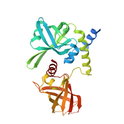
Crystal structure of the Borreliella burgdorferi PlzA protein in complex with c-di-GMP


Experimental Data Snapshot

  • Method: X-RAY DIFFRACTION
  • Resolution: 1.60 Å
  • R-Value Free: 
    0.218 (Depositor), 0.217 (DCC) 
  • R-Value Work: 
    0.191 (Depositor), 0.191 (DCC) 
  • R-Value Observed: 
    0.192 (Depositor) 

wwPDB Validation 3D Report Full Report

Validation slider image for 7MIE

Ligand Structure Quality Assessment 


This is version 1.2 of the entry. See complete history

Literature

High-resolution crystal structure of the Borreliella burgdorferi PlzA protein in complex with c-di-GMP: new insights into the interaction of c-di-GMP with the novel xPilZ domain.

Singh, A.Izac, J.R.Schuler, E.J.A.Patel, D.T.Davies, C.Marconi, R.T.

(2021) Pathog Dis 79

  • DOI: https://doi.org/10.1093/femspd/ftab030
  • Primary Citation Related Structures: 
    7MIE

  • PubMed Abstract: 

    In the tick-borne pathogens, Borreliella burgdorferi and Borrelia hermsii, c-di-GMP is produced by a single diguanylate cyclase (Rrp1). In these pathogens, the Plz proteins (PlzA, B and C) are the only c-di-GMP receptors identified to date and PlzA is the sole c-di-GMP receptor found in all Borreliella isolates. Bioinformatic analyses suggest that PlzA has a unique PilZN3-PilZ architecture with the relatively uncommon xPilZ domain. Here, we present the crystal structure of PlzA in complex with c-di-GMP (1.6 Å resolution). This is the first structure of a xPilz domain in complex with c-di-GMP to be determined. PlzA has a two-domain structure, where each domain comprises topologically equivalent PilZ domains with minimal sequence identity but remarkable structural similarity. The c-di-GMP binding site is formed by the linker connecting the two domains. While the structure of apo PlzA could not be determined, previous fluorescence resonance energy transfer data suggest that apo and holo forms of the protein are structurally distinct. The information obtained from this study will facilitate ongoing efforts to identify the molecular mechanisms of PlzA-mediated regulation in ticks and mammals.


  • Organizational Affiliation
    • Department of Biochemistry and Molecular Biology, Medical University of South Carolina, Charleston, South Carolina 29425, USA.

Macromolecule Content 

  • Total Structure Weight: 33.59 kDa 
  • Atom Count: 2,172 
  • Modeled Residue Count: 242 
  • Deposited Residue Count: 284 
  • Unique protein chains: 1

Macromolecules

Find similar proteins by:|  3D Structure
Entity ID: 1
MoleculeChains  Sequence LengthOrganismDetailsImage
PlzA284Borreliella burgdorferiMutation(s): 0 
Gene Names: BB_0733
UniProt
Find proteins for O51675 (Borreliella burgdorferi (strain ATCC 35210 / DSM 4680 / CIP 102532 / B31))
Explore O51675 
Go to UniProtKB:  O51675
Entity Groups
Sequence Clusters30% Identity50% Identity70% Identity90% Identity95% Identity100% Identity
UniProt GroupO51675
Sequence Annotations
Expand
Reference Sequence

Small Molecules

Ligands 4 Unique
IDChains Name / Formula / InChI Key2D Diagram3D Interactions
C2E
(Subject of Investigation/LOI)

Query on C2E


Download:Ideal Coordinates CCD File
B [auth A],
C [auth A]
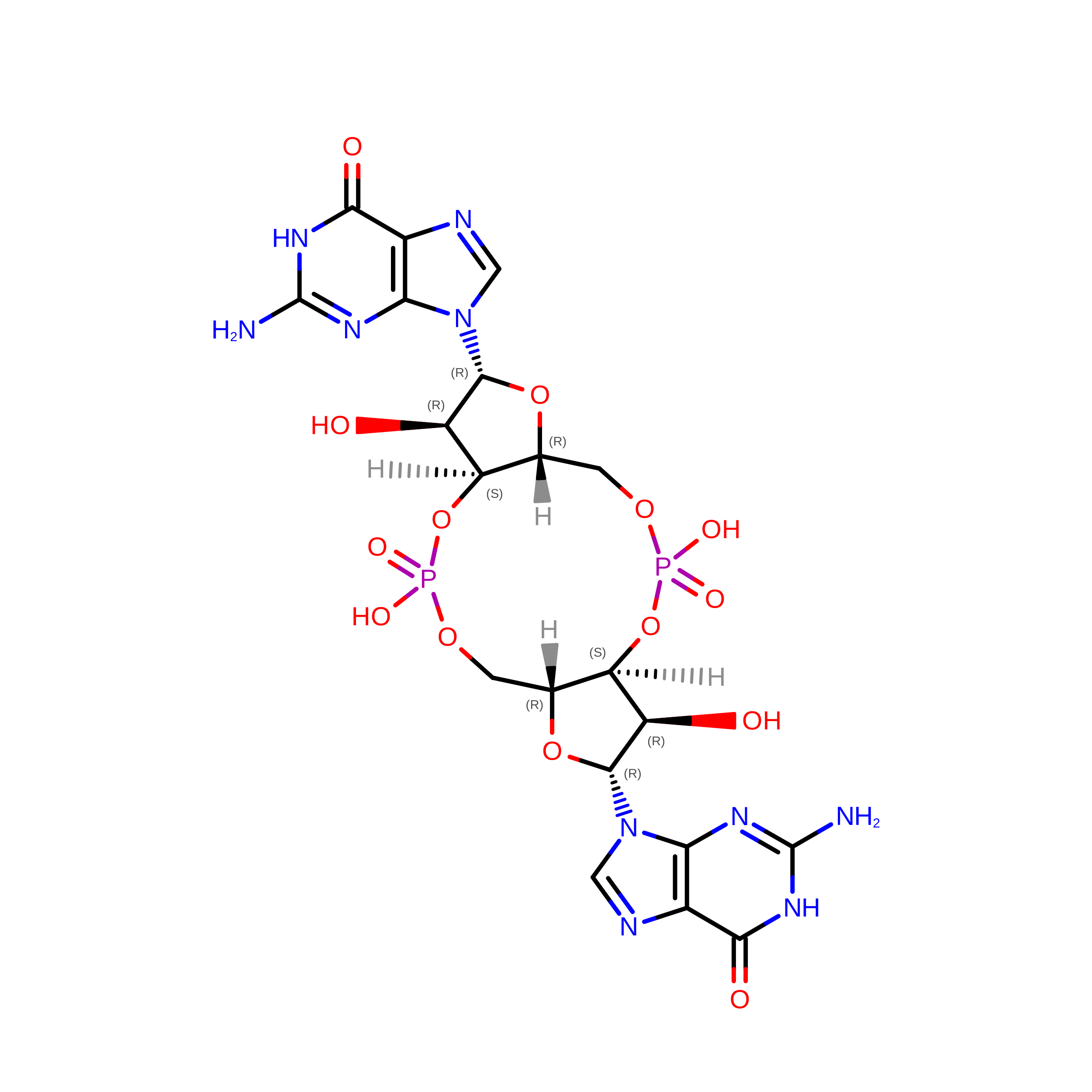
9,9'-[(2R,3R,3aS,5S,7aR,9R,10R,10aS,12S,14aR)-3,5,10,12-tetrahydroxy-5,12-dioxidooctahydro-2H,7H-difuro[3,2-d:3',2'-j][1,3,7,9,2,8]tetraoxadiphosphacyclododecine-2,9-diyl]bis(2-amino-1,9-dihydro-6H-purin-6-one)
C20 H24 N10 O14 P2
PKFDLKSEZWEFGL-MHARETSRSA-N
PEG

Query on PEG


Download:Ideal Coordinates CCD File
E [auth A]DI(HYDROXYETHYL)ETHER
C4 H10 O3
MTHSVFCYNBDYFN-UHFFFAOYSA-N
EDO

Query on EDO


Download:Ideal Coordinates CCD File
F [auth A],
G [auth A]
1,2-ETHANEDIOL
C2 H6 O2
LYCAIKOWRPUZTN-UHFFFAOYSA-N
CL

Query on CL


Download:Ideal Coordinates CCD File
D [auth A]CHLORIDE ION
Cl
VEXZGXHMUGYJMC-UHFFFAOYSA-M

Experimental Data & Validation

Experimental Data

  • Method: X-RAY DIFFRACTION
  • Resolution: 1.60 Å
  • R-Value Free:  0.218 (Depositor), 0.217 (DCC) 
  • R-Value Work:  0.191 (Depositor), 0.191 (DCC) 
  • R-Value Observed: 0.192 (Depositor) 
Space Group: P 1 21 1
Unit Cell:
Length ( Å )Angle ( ˚ )
a = 63.699α = 90
b = 36.861β = 113.9
c = 64.905γ = 90
Software Package:
Software NamePurpose
HKL-2000data reduction
HKL-2000data scaling
REFMACrefinement
PDB_EXTRACTdata extraction
PHENIXphasing

Structure Validation

View Full Validation Report


Ligand Structure Quality Assessment 


Entry History 

Deposition Data

Revision History  (Full details and data files)

  • Version 1.0: 2021-06-30
    Type: Initial release
  • Version 1.1: 2021-07-14
    Changes: Database references
  • Version 1.2: 2024-05-22
    Changes: Data collection, Database references