


Crystal structure of unknown function protein protein B9J08_000055 Candida auris
Chang, C., Evdokimova, E., Savchenko, A., Joachimiak, A., Center for Structural Genomics of Infectious Diseases (CSGID)To be published.
Experimental Data Snapshot
Starting Model: experimental
View more details
wwPDB Validation 3D Report Full Report

Entity ID: 1 | |||||
|---|---|---|---|---|---|
| Molecule | Chains | Sequence Length | Organism | Details | Image |
| Homoserine dehydrogenase | 359 | Candidozyma auris | Mutation(s): 1 Gene Names: B9J08_000055 EC: 1.1.1.3 | 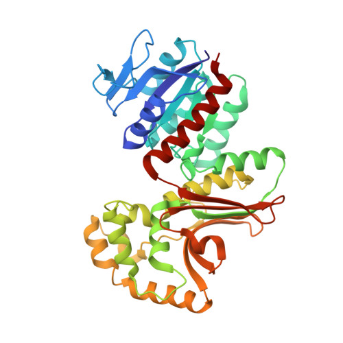 | |
UniProt | |||||
Entity Groups | |||||
| Sequence Clusters | 30% Identity50% Identity70% Identity90% Identity95% Identity100% Identity | ||||
| UniProt Group | A0A2H1A6V9 | ||||
Sequence AnnotationsExpand | |||||
Reference Sequence | |||||
| Length ( Å ) | Angle ( ˚ ) |
|---|---|
| a = 52.263 | α = 90 |
| b = 111.925 | β = 90 |
| c = 124.207 | γ = 90 |
| Software Name | Purpose |
|---|---|
| SCALEPACK | data scaling |
| SBC-Collect | data collection |
| PHENIX | refinement |
| PDB_EXTRACT | data extraction |
| HKL-3000 | data reduction |
| MOLREP | phasing |


