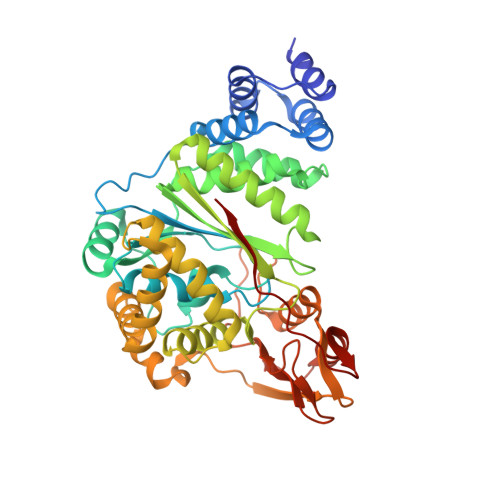
7M7K

Crystal structure of uridine bound to Geobacillus thermoglucosidasius pyrimidine nucleoside phosphorylase PyNP


Experimental Data Snapshot

  • Method: X-RAY DIFFRACTION
  • Resolution: 1.89 Å
  • R-Value Free: 0.208 
  • R-Value Work: 0.174 
  • R-Value Observed: 0.175 

wwPDB Validation   3D Report Full Report


Ligand Structure Quality Assessment 


This is version 1.1 of the entry. See complete history


Literature

Diversification of 4'-Methylated Nucleosides by Nucleoside Phosphorylases

Kaspar, F.Seeger, M.Westarp, S.Kollmann, C.Lehmann, A.P.Pausch, P.Kemper, S.Neubauer, P.Bange, G.Schallmey, A.Werz, D.B.Kurreck, A.

(2021) ACS Catal : 10830-10835


Macromolecules
Find similar proteins by:  (by identity cutoff)  |  3D Structure
Entity ID: 1
MoleculeChains Sequence LengthOrganismDetailsImage
Pyrimidine-nucleoside phosphorylaseA [auth B]438Parageobacillus thermoglucosidasiusMutation(s): 0 
Gene Names: DV714_16680
EC: 2.4.2.2
Entity Groups  
Sequence Clusters30% Identity50% Identity70% Identity90% Identity95% Identity100% Identity
Sequence Annotations
Expand
  • Reference Sequence
Experimental Data & Validation

Experimental Data

  • Method: X-RAY DIFFRACTION
  • Resolution: 1.89 Å
  • R-Value Free: 0.208 
  • R-Value Work: 0.174 
  • R-Value Observed: 0.175 
  • Space Group: P 41 21 2
Unit Cell:
Length ( Å )Angle ( ˚ )
a = 84.262α = 90
b = 84.262β = 90
c = 121.694γ = 90
Software Package:
Software NamePurpose
PHENIXrefinement
PHENIXrefinement
XDSdata reduction
Aimlessdata scaling
MOLREPphasing

Structure Validation

View Full Validation Report


Ligand Structure Quality Assessment 


Entry History 

Deposition Data

Revision History  (Full details and data files)

  • Version 1.0: 2021-08-25
    Type: Initial release
  • Version 1.1: 2023-10-18
    Changes: Data collection, Refinement description