7M7A | pdb_00007m7a

Legionella pneumophila MavQ lipid kinase

  • Classification: TRANSFERASE
  • Organism(s): Legionella pneumophila
  • Expression System: Escherichia coli BL21(DE3)
  • Mutation(s): No 

  • Deposited: 2021-03-27 Released: 2021-04-28 
  • Deposition Author(s): Tomchick, D.R., Tagliabracci, V.S., Hsieh, T.S., Lopez, V.A.
  • Funding Organization(s): National Institutes of Health/National Institute of General Medical Sciences (NIH/NIGMS), National Institutes of Health/National Heart, Lung, and Blood Institute (NIH/NHLBI), National Institutes of Health/Office of the Director, Robert A. Welch Foundation, Cancer Prevention and Research Institute of Texas (CPRIT), National Center for Research and Development (Poland)

Experimental Data Snapshot

  • Method: X-RAY DIFFRACTION
  • Resolution: 3.20 Å
  • R-Value Free: 
    0.283 (Depositor), 0.278 (DCC) 
  • R-Value Work: 
    0.239 (Depositor), 0.238 (DCC) 
  • R-Value Observed: 
    0.241 (Depositor) 

wwPDB Validation 3D Report Full Report

Validation slider image for 7M7A

Ligand Structure Quality Assessment 


This is version 1.3 of the entry. See complete history

Literature

Dynamic remodeling of host membranes by self-organizing bacterial effectors.

Hsieh, T.S.Lopez, V.A.Black, M.H.Osinski, A.Pawlowski, K.Tomchick, D.R.Liou, J.Tagliabracci, V.S.

(2021) Science 372: 935-941

  • DOI: https://doi.org/10.1126/science.aay8118
  • Primary Citation Related Structures: 
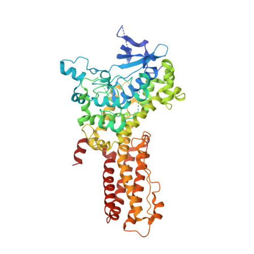
    7M7A

  • PubMed Abstract: 

    During infection, intracellular bacterial pathogens translocate a variety of effectors into host cells that modify host membrane trafficking for their benefit. We found a self-organizing system consisting of a bacterial phosphoinositide kinase and its opposing phosphatase that formed spatiotemporal patterns, including traveling waves, to remodel host cellular membranes. The Legionella effector MavQ, a phosphatidylinositol (PI) 3-kinase, was targeted to the endoplasmic reticulum (ER). MavQ and the Legionella PI 3-phosphatase SidP, even in the absence of other bacterial components, drove rapid PI 3-phosphate turnover on the ER and spontaneously formed traveling waves that spread along ER subdomains inducing vesicle and tubule budding. Thus, bacteria can exploit a self-organizing membrane-targeting mechanism to hijack host cellular structures for survival.


  • Organizational Affiliation
    • Department of Molecular Biology, University of Texas Southwestern Medical Center, Dallas, TX 75390, USA.

Macromolecule Content 

  • Total Structure Weight: 275.72 kDa 
  • Atom Count: 17,652 
  • Modeled Residue Count: 2,125 
  • Deposited Residue Count: 2,360 
  • Unique protein chains: 1

Macromolecules

Find similar proteins by:|  3D Structure
Entity ID: 1
MoleculeChains  Sequence LengthOrganismDetailsImage
Phosphoinositide 3-kinase MavQ
A, B, C, D
590Legionella pneumophilaMutation(s): 0 
Gene Names: lpg2975
EC: 3.1.3.64
UniProt
Find proteins for Q5ZRA8 (Legionella pneumophila subsp. pneumophila (strain Philadelphia 1 / ATCC 33152 / DSM 7513))
Explore Q5ZRA8 
Go to UniProtKB:  Q5ZRA8
Entity Groups
Sequence Clusters30% Identity50% Identity70% Identity90% Identity95% Identity100% Identity
UniProt GroupQ5ZRA8
Sequence Annotations
Expand
Reference Sequence

Small Molecules

Ligands 2 Unique
IDChains Name / Formula / InChI Key2D Diagram3D Interactions
ADP
(Subject of Investigation/LOI)

Query on ADP


Download:Ideal Coordinates CCD File
E [auth A],
G [auth B],
I [auth C],
K [auth D]
ADENOSINE-5'-DIPHOSPHATE
C10 H15 N5 O10 P2
XTWYTFMLZFPYCI-KQYNXXCUSA-N
MG
(Subject of Investigation/LOI)

Query on MG


Download:Ideal Coordinates CCD File
F [auth A],
H [auth B],
J [auth C],
L [auth D]
MAGNESIUM ION
Mg
JLVVSXFLKOJNIY-UHFFFAOYSA-N

Experimental Data & Validation

Experimental Data

  • Method: X-RAY DIFFRACTION
  • Resolution: 3.20 Å
  • R-Value Free:  0.283 (Depositor), 0.278 (DCC) 
  • R-Value Work:  0.239 (Depositor), 0.238 (DCC) 
  • R-Value Observed: 0.241 (Depositor) 
Space Group: C 2 2 21
Unit Cell:
Length ( Å )Angle ( ˚ )
a = 69.399α = 90
b = 210.235β = 90
c = 499.861γ = 90
Software Package:
Software NamePurpose
PHENIXrefinement
HKL-3000data scaling
HKL-3000data reduction
PDB_EXTRACTdata extraction
SHELXDphasing
MLPHAREphasing
PARROTphasing
BUCCANEERmodel building
Cootmodel building

Structure Validation

View Full Validation Report


Ligand Structure Quality Assessment 


Entry History 

& Funding Information

Deposition Data


Funding OrganizationLocationGrant Number
National Institutes of Health/National Institute of General Medical Sciences (NIH/NIGMS)United StatesDP2GM137419
National Institutes of Health/National Institute of General Medical Sciences (NIH/NIGMS)United StatesR01GM113079
National Institutes of Health/National Institute of General Medical Sciences (NIH/NIGMS)United StatesT32GM008203-29
National Institutes of Health/National Heart, Lung, and Blood Institute (NIH/NHLBI)United StatesF30HL143859-01
National Institutes of Health/Office of the DirectorUnited StatesS10OD025018
Robert A. Welch FoundationUnited StatesI-1911
Robert A. Welch FoundationUnited StatesI-1789
Cancer Prevention and Research Institute of Texas (CPRIT)United StatesRP170674
National Center for Research and Development (Poland)PolandPPN/BEK/2018/1/00431
National Institutes of Health/Office of the DirectorUnited StatesS10OD021685

Revision History  (Full details and data files)

  • Version 1.0: 2021-04-28
    Type: Initial release
  • Version 1.1: 2021-05-12
    Changes: Database references
  • Version 1.2: 2021-06-09
    Changes: Database references
  • Version 1.3: 2024-05-22
    Changes: Data collection, Database references