7LXJ

Bacillus cereus DNA glycosylase AlkD bound to a duocarmycin SA-adenine nucleobase adduct and DNA containing an abasic site


Experimental Data Snapshot

  • Method: X-RAY DIFFRACTION
  • Resolution: 1.93 Å
  • R-Value Free: 0.201 
  • R-Value Work: 0.154 
  • R-Value Observed: 0.156 

wwPDB Validation   3D Report Full Report


Ligand Structure Quality Assessment 


This is version 1.2 of the entry. See complete history


Literature

Structural evolution of a DNA repair self-resistance mechanism targeting genotoxic secondary metabolites.

Mullins, E.A.Dorival, J.Tang, G.L.Boger, D.L.Eichman, B.F.

(2021) Nat Commun 12: 6942-6942

  • DOI: https://doi.org/10.1038/s41467-021-27284-7
  • Primary Citation of Related Structures:  
    7LXH, 7LXJ

  • PubMed Abstract: 

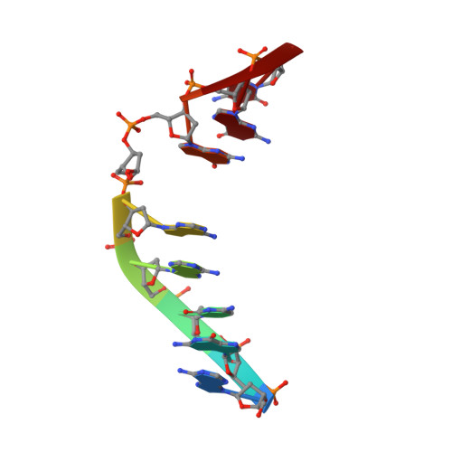
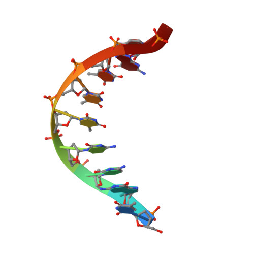
    Microbes produce a broad spectrum of antibiotic natural products, including many DNA-damaging genotoxins. Among the most potent of these are DNA alkylating agents in the spirocyclopropylcyclohexadienone (SCPCHD) family, which includes the duocarmycins, CC-1065, gilvusmycin, and yatakemycin. The yatakemycin biosynthesis cluster in Streptomyces sp. TP-A0356 contains an AlkD-related DNA glycosylase, YtkR2, that serves as a self-resistance mechanism against yatakemycin toxicity. We previously reported that AlkD, which is not present in an SCPCHD producer, provides only limited resistance against yatakemycin. We now show that YtkR2 and C10R5, a previously uncharacterized homolog found in the CC-1065 biosynthetic gene cluster of Streptomyces zelensis, confer far greater resistance against their respective SCPCHD natural products. We identify a structural basis for substrate specificity across gene clusters and show a correlation between in vivo resistance and in vitro enzymatic activity indicating that reduced product affinity-not enhanced substrate recognition-is the evolutionary outcome of selective pressure to provide self-resistance against yatakemycin and CC-1065.


  • Organizational Affiliation

    Department of Biological Sciences, Vanderbilt University, Nashville, Tennessee, USA.


Macromolecules

Find similar proteins by:  (by identity cutoff)  |  3D Structure
Entity ID: 1
MoleculeChains Sequence LengthOrganismDetailsImage
DNA-7-methylguanine glycosylase241Bacillus cereusMutation(s): 0 
Gene Names: bcere0015_46090
UniProt
Find proteins for Q816E8 (Bacillus cereus (strain ATCC 14579 / DSM 31 / CCUG 7414 / JCM 2152 / NBRC 15305 / NCIMB 9373 / NCTC 2599 / NRRL B-3711))
Explore Q816E8 
Go to UniProtKB:  Q816E8
Entity Groups  
Sequence Clusters30% Identity50% Identity70% Identity90% Identity95% Identity100% Identity
UniProt GroupQ816E8
Sequence Annotations
Expand
  • Reference Sequence

Find similar nucleic acids by:  Sequence   |   3D Structure  

Entity ID: 2
MoleculeChains LengthOrganismImage
DNA (5'-D(*AP*GP*CP*AP*AP*(ORP)P*GP*GP*C)-3')9synthetic construct
Sequence Annotations
Expand
  • Reference Sequence

Find similar nucleic acids by:  Sequence   |   3D Structure  

Entity ID: 3
MoleculeChains LengthOrganismImage
DNA (5'-D(*TP*GP*CP*CP*TP*TP*TP*GP*C)-3')9synthetic construct
Sequence Annotations
Expand
  • Reference Sequence
Small Molecules
Ligands 2 Unique
IDChains Name / Formula / InChI Key2D Diagram3D Interactions
YNY (Subject of Investigation/LOI)
Query on YNY

Download Ideal Coordinates CCD File 
F [auth B]methyl (8R)-8-{[(4P)-6-amino-3H-purin-3-yl]methyl}-4-hydroxy-6-(5,6,7-trimethoxy-1H-indole-2-carbonyl)-3,6,7,8-tetrahydropyrrolo[3,2-e]indole-2-carboxylate
C30 H28 N8 O7
AZHBDEJKEVQJNC-CQSZACIVSA-N
CA
Query on CA

Download Ideal Coordinates CCD File 
D [auth A],
E [auth B]
CALCIUM ION
Ca
BHPQYMZQTOCNFJ-UHFFFAOYSA-N
Experimental Data & Validation

Experimental Data

  • Method: X-RAY DIFFRACTION
  • Resolution: 1.93 Å
  • R-Value Free: 0.201 
  • R-Value Work: 0.154 
  • R-Value Observed: 0.156 
  • Space Group: C 1 2 1
Unit Cell:
Length ( Å )Angle ( ˚ )
a = 124.715α = 90
b = 55.661β = 112.2
c = 47.991γ = 90
Software Package:
Software NamePurpose
HKL-2000data reduction
DENZOdata reduction
HKL-2000data scaling
SCALEPACKdata scaling
PHENIXphasing
PHASERphasing
Cootmodel building
PHENIXrefinement
PDB_EXTRACTdata extraction

Structure Validation

View Full Validation Report


Ligand Structure Quality Assessment 


Entry History & Funding Information

Deposition Data


Funding OrganizationLocationGrant Number
National Science Foundation (NSF, United States)United StatesMCB-1928918

Revision History  (Full details and data files)

  • Version 1.0: 2021-11-17
    Type: Initial release
  • Version 1.1: 2021-12-08
    Changes: Database references
  • Version 1.2: 2023-10-18
    Changes: Data collection, Refinement description